Revolutionary AI Transforms Tissue Analysis with Virtual Staining
2025-07-01
Author: Siti
In the realm of disease diagnosis, traditional histopathology relies heavily on the meticulous process of chemically staining tissue samples. This method, often cumbersome and destructive, involves using harmful dyes to enhance visibility under a microscope—making it not only time-consuming but also costly.
However, a groundbreaking innovation is reshaping this field: the advent of "virtual staining." Researchers at the University of California, Los Angeles (UCLA) have unveiled an AI-driven tool that can transform unstained tissue images into vibrant, detail-rich representations without any chemical interventions.
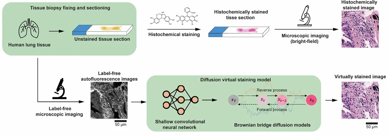
Published in Nature Communications, the study highlights a revolutionary approach using a diffusion model inspired by Brownian motion. This new AI technique generates exceptionally high-resolution microscopic images, effectively stepping in for traditional histochemical staining and offering a cost-efficient, non-destructive alternative.
The virtual staining method boasts the remarkable ability to enhance low-resolution autofluorescence images into stunning, high-fidelity brightfield images, mimicking the appearance of commonly used stains like hematoxylin and eosin (H&E). With remarkable precision, this technique achieves a spatial resolution increase of four to five times, significantly improving both visual presentation and diagnostic capability.
One of the standout features of this research is its innovative control over the randomness associated with diffusion models. Employing advanced strategies for sampling, the researchers reduced inconsistencies between images, ensuring reliable and uniform results vital for clinical diagnostics.
Senior author Professor Aydogan Ozcan states, "Diffusion models are incredibly powerful, but their randomness can be unpredictable. We developed a method to harness that unpredictability, giving us the control we need for consistent outcomes—critical for real-world medical applications."
In blind tests using human lung tissue samples, this AI model outperformed existing methods, achieving superior resolution and accuracy. A certified pathologist verified the alignment between AI-generated images and their chemically stained equivalents, solidifying the technology’s reliability.
The effectiveness of this virtual staining approach was further confirmed when applied to human heart tissue, demonstrating high precision across diverse types. By eliminating the need for chemical staining, this innovation not only conserves resources and time but also safeguards the integrity of the tissue being analyzed.
This pioneering technology could significantly enhance digital pathology processes, especially in environments with limited resources or within urgent clinical scenarios.
By uniting pixel super-resolution with virtual staining, this AI initiative ushers in a new era for digital pathology, inching us closer to the promise of precision medicine without the complexities of traditional staining techniques. This research marks a pivotal moment in computational pathology, establishing a new benchmark for consistent, high-quality virtual staining of unmarked tissues.


 Brasil (PT)
 Canada (EN)
 Chile (ES)
 Česko (CS)
 대한민국 (KO)
 España (ES)
 France (FR)
 Hong Kong (EN)
 Italia (IT)
 日本 (JA)
 Magyarország (HU)
 Norge (NO)
 Polska (PL)
 Schweiz (DE)
 Singapore (EN)
 Sverige (SV)
 Suomi (FI)
 Türkiye (TR)
 الإمارات العربية المتحدة (AR)