Revolutionizing Tissue Imaging: AI Transforms Mass Spectrometry into Virtual Histology
2025-08-04
Author: Michael
A Breakthrough in Imaging Technology
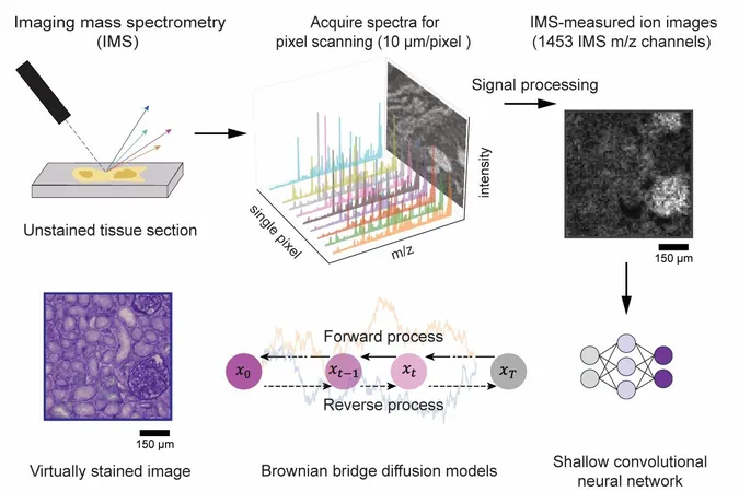
An exciting international collaboration between researchers from UCLA, Vanderbilt University, and Delft University of Technology has announced a groundbreaking advancement in biological research: an artificial intelligence (AI) method that virtually 'stains' images derived from imaging mass spectrometry (IMS)!
Enhanced Resolution, No Chemicals Required
Published in the esteemed journal *Science Advances*, this innovative technique significantly boosts spatial resolution and reveals intricate cellular details—yet it does so without any chemical staining. By utilizing a cutting-edge diffusion-based generative model, researchers have managed to create digital images that rival traditional histochemical stains, all while preserving precious tissue samples.
Unlocking the Power of Imaging Mass Spectrometry
IMS is a robust tool known for its ability to map a vast array of molecular species in biological tissues with remarkable chemical specificity. However, conventional IMS has a notable limitation: relatively low spatial resolution and a lack of cellular detail, making it challenging to glean critical insights into tissue structure.
A New Era of Virtual Staining
To tackle these limitations, the research team developed a novel virtual staining approach. This method electronically transforms lower-resolution, label-free IMS data into high-resolution brightfield microscopy images—effectively mimicking the appearance of tissues stained with the Periodic Acid–Schiff (PAS) method, which highlights vital polysaccharides and glycoproteins.
AI Surpasses Traditional Imaging Limitations
What’s truly astounding is that this AI framework achieves such high-quality results even though the pixel size in IMS data is nearly ten times larger than those of traditional optical microscopy images.
Bridging Molecular and Cellular Insights
Professor Aydogan Ozcan of UCLA, the study's lead author, enthusiastically stated, "This diffusion-based approach dramatically enhances the interpretability of mass spectrometry images, bridging the gap between molecular specificity and cellular morphology—all without the need for chemical staining!"
Real-World Applications and Reliability
In blind tests conducted with human kidney tissues, the virtually stained images showed remarkable alignment with chemically stained counterparts, allowing pathologists to accurately identify critical structures and disease markers using just the virtual images.
Streamlining Biomedical Research
This revolutionary technique holds tremendous promise for IMS-driven biomedical research and diagnostics. By removing the necessity for cumbersome chemical staining and complex imaging processes, it preserves tissue integrity for subsequent molecular analyses, vastly improving efficiency in mass spectrometry-based workflows.
A Vision for the Future of Diagnostics
Professor Ozcan concluded with an optimistic outlook: "We believe this method will pave the way for exciting new advancements in spatial biology and clinical diagnostics. By digitally generating high-quality histological images from mass spectrometry data, we can streamline workflows and potentially foster monumental strides in biomedical discovery."


 Brasil (PT)
 Canada (EN)
 Chile (ES)
 Česko (CS)
 대한민국 (KO)
 España (ES)
 France (FR)
 Hong Kong (EN)
 Italia (IT)
 日本 (JA)
 Magyarország (HU)
 Norge (NO)
 Polska (PL)
 Schweiz (DE)
 Singapore (EN)
 Sverige (SV)
 Suomi (FI)
 Türkiye (TR)
 الإمارات العربية المتحدة (AR)