Unveiling Aquatic Life: Free Video Microscopy Transforms Our Understanding of Microbial Behavior in Extreme Environments
2025-04-04
Author: Sarah
Groundbreaking Investigation
A groundbreaking investigation has utilized in situ video microscopy to explore the active motion, morphology, and optical properties of microbes as potential biosignatures in extreme aquatic environments—places that have been largely overlooked in past studies.
Challenging Field Sites
This pioneering research spanned a variety of challenging field sites, including samples of seawater, brines from sea ice, cryopeg brines, hypersaline pools, hyperalkaline springs, and glaciovolcanic cave ice. Remarkably, active microbial motion was detected in all samples, except for cryopeg brine, unless the samples were subjected to a temperature gradient above their in situ conditions. The evidence of life was captured through impressive video microscopy techniques.
Fascinating Discoveries
Images captured reveal fascinating details, such as an amplitude image of a non-motile diatom from Greenland's sea ice brine, showcasing clear cell wall and organelle boundaries. More significantly, rapidly swimming organisms were recorded, providing insights into the micro-ecosystems thriving under frozen environments. Remarkably, motile tracks of small microbes were captured from Greenland seawater after being incubated overnight, illustrating the intricate behaviors of these life forms.
Understanding Microbial Behavior
An intriguing discovery was that motility levels were generally low at temperatures below 4°C. However, researchers could differentiate non-motile cells from microminerals based on passive motion, refractive indexes, and morphology of larger eukaryotic cells. This opens new avenues for understanding how life adapts to extreme cold.
Clever Experimental Setups
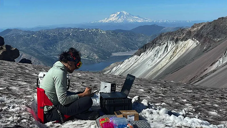
The research also highlighted clever experimental setups in various unique locations, such as sea ice brine sackholes in Nuuk, Greenland, and permafrost tunnels in Utqiaġvik, Alaska, which allowed for extensive data collection on microbial behavior under ambient conditions.
Environmental Stimuli Effects
Strikingly, simple environmental stimuli like warming or the introduction of L-serine significantly increased the motile fraction of cells. Instances of chemotaxis and thermotaxis were also documented, revealing the sophisticated responses of these microorganisms to their surroundings. Utilizing an open-source, autonomous software package that can function on scaled-down spaceflight computers, researchers effectively classified vast amounts of data from these experiments.
Implications for Astrobiology
These findings underscore the necessity of volumetric light microscopy for astrobiological research, particularly for life detection missions, while also emphasizing the importance of developing methods to stimulate microbial activity in situ. This is especially critical when considering the limitations imposed by mission bandwidth and the necessity for instruments capable of capturing and analyzing cell-like objects in detail.
Visualizations of Microbial Interactions
In addition to measuring motion in these extreme environments, the research showcased impressive visualizations of large microbes and their interactions, revealing complex trajectories and potential predatory behaviors.
Significance of the Study
This pivotal study not only enhances our understanding of life in remote aquatic environments on Earth but also has significant implications for astrobiology, shedding light on potential life forms that could exist beyond our planet. As scientists continue to explore these extraordinary ecosystems, the implications for future space missions in the search for extraterrestrial life remain expansive and exciting.
Conclusion
Stay tuned as we continue to unravel the mysteries of life in extreme environments and look forward to groundbreaking discoveries that could redefine how we view life in the universe!


 Brasil (PT)
 Canada (EN)
 Chile (ES)
 Česko (CS)
 대한민국 (KO)
 España (ES)
 France (FR)
 Hong Kong (EN)
 Italia (IT)
 日本 (JA)
 Magyarország (HU)
 Norge (NO)
 Polska (PL)
 Schweiz (DE)
 Singapore (EN)
 Sverige (SV)
 Suomi (FI)
 Türkiye (TR)
 الإمارات العربية المتحدة (AR)