Unlocking Brain Mysteries: Groundbreaking Research Reveals Hidden Signals in Brain Imaging Technology
2024-11-11
Author: Arjun
In an exciting breakthrough for neuroscience, researchers are delving deep into the intricate workings of the brain using a pioneering technique known as resting-state functional magnetic resonance imaging (rsfMRI). This cutting-edge method measures brain activity by tracking alterations in blood flow to various regions of the brain, yet it has long been unclear how these blood flow variations correspond to the underlying activity of neurons—the essential cells that send and receive messages through electrical signals.
Understanding rsfMRI: The Current Landscape and Its Limitations
Resting-state fMRI allows researchers to observe the collaborative activity of different brain regions by measuring spontaneous alterations in blood flow—establishing what are known as resting-state brain networks (RSNs). Despite its widespread use, the definitive connection between these blood flow changes and actual neural excitement remains murky, underscoring the need for further investigation.
To tackle this issue, Zhang’s research incorporates synchronizing rsfMRI with electrophysiology signals—an approach that captures both dynamic blood flow and direct neural activity from the same brain area simultaneously. This simultaneous recording aims to shed light on the relationship between spontaneous blood flow fluctuations and neuronal firing, creating a clearer picture of brain operations.
Revolutionary Findings: A New Perspective on Brain Signals
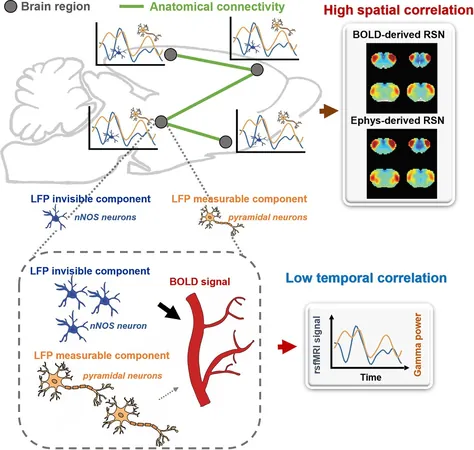
The research unveiled a surprising disparity in the spatial and temporal dynamics between electrophysiology signals, which directly measure neural firing, and the rsfMRI signals representative of blood flow. While the structural patterns of brain-wide RSN connectivity detected through rsfMRI matched those from electrophysiological data, the timing of these signals did not align, suggesting complexities in brain activity that previous models have not accounted for.
This revelation posits the existence of what the researchers term "invisible signals" in electrophysiology—silent contributors to rsfMRI signals that extend our understanding of functional brain networks. Traditionally, it has been assumed that electrophysiological signals primarily drive rsfMRI signals; however, this study indicates that significant portions of rsfMRI data may arise from these non-visible components.
Implications for Future Research and Human Studies
These findings challenge established theories about how rsfMRI signals are interpreted, highlighting the necessity for deeper insights into the neural foundations that shape these signals. The research underscores that if RSNs can be influenced significantly by unobservable neural mechanisms, our interpretations of brain imaging data might need fundamental reassessment.
The implications of this study extend beyond animal models. The mechanisms governing rsfMRI in mice are likely applicable to human brains, allowing researchers to harness these findings for future studies concerning human brain function and disorders. By further investigating this underexplored territory, scientists hope to unlock new avenues for understanding various neurological conditions and enhance the efficacy of brain imaging techniques.
This exciting research not only augments our grasp of brain activity but also paves the way for future innovations in medical imaging and neuroscience, potentially revolutionizing our approach to understanding the human brain.


 Brasil (PT)
 Canada (EN)
 Chile (ES)
 España (ES)
 France (FR)
 Hong Kong (EN)
 Italia (IT)
 日本 (JA)
 Magyarország (HU)
 Norge (NO)
 Polska (PL)
 Schweiz (DE)
 Singapore (EN)
 Sverige (SV)
 Suomi (FI)
 Türkiye (TR)