Revolutionary Tricorder Tech: New Imaging Technique Unlocks Hidden Biomolecules in Cells
2025-04-27
Author: Mei
Discovering the Invisible: A Game-Changer in Biomolecular Analysis
As humanity forges ahead into the cosmos, advanced astrobiology probes will require cutting-edge technology to analyze alien worlds in real-time—without human intervention. When we finally send crews to explore these distant realms, maximizing their limited study time becomes paramount. Recent breakthroughs in imaging technology promise to revolutionize our understanding of extraterrestrial ecosystems.
The Challenge of Visualization in Biological Research
In the world of biology, the adage "seeing is believing" holds true. Yet, researchers often grapple with the limitations of existing imaging techniques. The quest to simultaneously visualize the myriad biomolecules within a single, intact tissue sample remains a daunting challenge. Understanding the location and interaction of hundreds of biomolecules—from lipids to proteins—inside their natural habitats is crucial for unraveling their functions and contributions.
Traditional Techniques Fall Short
While imaging methods like microscopy provide essential insights, they typically track only a limited number of molecules at once and miss many types, including various lipids. Conventional mass spectrometry excels by identifying hundreds of biomolecules but falls short as it requires samples to be destroyed, leaving researchers blind to the spatial arrangements of these vital components.
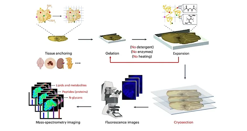
A Breakthrough Method: Expansion Mass Spectrometry Imaging (TEMI)
Enter the innovative expansion microscopy technique, co-invented by Paul Tillberg during his time at MIT. This groundbreaking approach employs a swellable hydrogel that uniformly expands biological samples, allowing scientists to observe finer details with standard microscopes. Now, this decade-old method has been adapted to enhance mass spectrometry imaging capabilities.
By gradually expanding intact tissues without compromising molecular integrity, researchers can now utilize mass spectrometry imaging to detect hundreds of molecules at the single-cell level within their native environments. This breakthrough provides unprecedented insights into molecular landscapes.
Exploring Cerebellum Patterns: New Discoveries Unveiled
Using this transformative technique, scientists mapped specific spatial patterns of small molecules across different layers of the cerebellum. Astonishingly, they discovered that biomolecules—including lipids, peptides, and proteins—are not uniformly distributed as once believed; each layer possesses its unique molecular signature.
Adapting the Technique for Broad Applications
The implications of this method extend beyond the cerebellum. Successful applications have been reported in various tissues, including kidney, pancreas, and tumor samples. In particular, the team revealed striking variations in biomolecules within tumor tissues, holding promise for deciphering tumor biology and aiding drug development.
A Hope for the Future: Accessibility and Adoption
By creating a technique that doesn’t require specialized equipment, the research team hopes to democratize this powerful imaging method for labs globally. The ease of learning the expansion technique is designed to make mass spectrometry imaging a staple tool for biologists everywhere. As mentioned by Wang, their goal was to develop a system that can be easily adopted, fostering broader advances in the field.
The New Frontier in Biomolecular Research
As this new technique, termed Tissue-Expansion Mass-Spectrometry Imaging (TEMI), gains traction, it could rewrite the rules of biomolecular exploration, paving the way for deeper insights into aging, disease, and even the molecular signatures of alien life forms. The ability to visualize biomolecules in their natural state could change everything we know about how life operates at the molecular level.


 Brasil (PT)
 Canada (EN)
 Chile (ES)
 Česko (CS)
 대한민국 (KO)
 España (ES)
 France (FR)
 Hong Kong (EN)
 Italia (IT)
 日本 (JA)
 Magyarország (HU)
 Norge (NO)
 Polska (PL)
 Schweiz (DE)
 Singapore (EN)
 Sverige (SV)
 Suomi (FI)
 Türkiye (TR)
 الإمارات العربية المتحدة (AR)