
Revolutionary Breakthrough: Chinese Scientists Create Miniature Microscope for Deep Brain Imaging
2025-08-23
Author: Jia
A New Era in Brain Research
In an astonishing leap for neuroscience, Chinese scientists have pioneered the world’s first high-resolution, multicolor deep-brain imaging technique in freely moving mice. This groundbreaking work was made possible through a state-of-the-art miniature two-photon microscope, as reported in the prestigious journal, Nature Methods.
The Challenge of Understanding the Brain
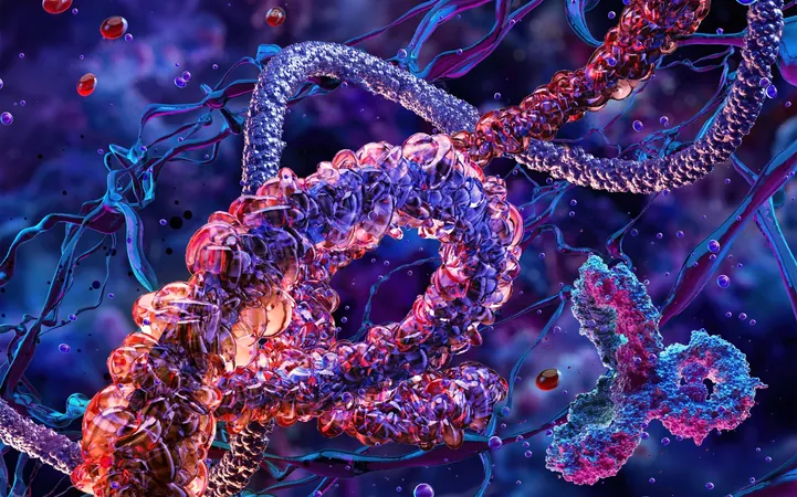
For decades, capturing the intricate dance between billions of neurons and trillions of synapses has posed a formidable challenge for scientists. The brain's functionality relies on this complex interplay, making real-time observation essential for decoding its mechanisms.
What is Two-Photon Microscopy?
Two-photon microscopy is a nonlinear optical imaging technique that provides remarkable resolution and the ability to visualize structures deep within biological tissues. It represents a significant advancement over traditional imaging methods.
The Journey to Innovation
In 2017, a trailblazing team from Peking University, led by Cheng Heping, unveiled the first generation of miniature two-photon microscopes. This achievement enabled researchers to achieve clear synaptic imaging in moving mice, marking a significant milestone in neuroscience.
The Breakthrough Fiber Technology
A key innovation in this latest research is the development of a new ultra-broadband hollow-core fiber, allowing the transmission of femtosecond pulsed lasers across a spectrum of wavelengths from 700 to 1,060 nanometers. This advancement overcomes previous limitations and introduces multicolor imaging capabilities in a lightweight, 2.6-gram device.
Unveiling Alzheimer's Disease
By applying this innovative microscope to mice with Alzheimer's, researchers succeeded in capturing vibrant three-color images representing various neuronal activities—red for calcium signals, green for mitochondrial interactions, and blue for plaque deposits. This provides unprecedented insights into cellular behavior and mitochondrial activity associated with early-stage Alzheimer's pathology.
A Live Broadcast of Brain Activity
Wu Runlong, one of the leading researchers, described the technology as a 'live color broadcast' of neuronal and organelle dynamics. By labeling different cell types with specific fluorescent markers, this breakthrough allows scientists to visualize complex interactions and networks within the brain.
Deeper Insights, Greater Depths
Remarkably, this advanced microscope can obtain neuronal data and structural images at depths exceeding 820 micrometers—currently the deepest observed without causing any tissue damage.
Versatile Imaging Capabilities
Furthermore, researchers can effortlessly switch between large-field and high-resolution imaging modes with simple adjustments. This adaptability allows them to capture a detailed close-up or a broader panoramic view of brain activity.
A Blueprint for Future Research
This monumental advancement in multicolor, deep-brain imaging positions scientists at the forefront of understanding cognitive processes, unraveling brain disease mechanisms, and exploring neuropharmaceutical effects. It heralds an exciting frontier in brain-computer interface development, making this technology a potential game-changer in neuroscience.


 Brasil (PT)
 Canada (EN)
 Chile (ES)
 Česko (CS)
 대한민국 (KO)
 España (ES)
 France (FR)
 Hong Kong (EN)
 Italia (IT)
 日本 (JA)
 Magyarország (HU)
 Norge (NO)
 Polska (PL)
 Schweiz (DE)
 Singapore (EN)
 Sverige (SV)
 Suomi (FI)
 Türkiye (TR)
 الإمارات العربية المتحدة (AR)