Revolutionary AI Tool Transforms Medical Imaging with Minimal Data
2025-08-01
Author: Arjun
Game-Changer in Medical Imaging
A groundbreaking artificial intelligence (AI) tool is set to revolutionize how doctors and researchers approach medical imaging, making the process faster and more cost-effective—even when only a few patient scans are available.
A Leap in Image Segmentation
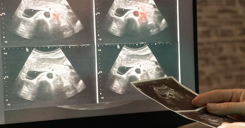
This innovative AI advances the technique known as medical image segmentation, where every pixel in an image is tagged to indicate its relevance—whether it's cancerous tissue or healthy. Traditionally, this painstaking task is carried out by highly trained specialists. While deep learning has shown promise in automating the segmentation process, it has faced a significant hurdle: the need for vast amounts of annotated images.
Breaking the Data Barrier
Li Zhang, a Ph.D. student at UC San Diego, explains that deep learning methods require large datasets that are often impractically costly and time-consuming to create. Many medical conditions lack the substantial data necessary for such advanced techniques.
To tackle this issue, Zhang and a dedicated team, led by Professor Pengtao Xie, have developed an AI model capable of learning image segmentation from minimal expert-labeled samples. This revolutionary tool cuts down the data needed by up to 20 times, paving the way for quicker and cheaper diagnostic tools, particularly in resource-strapped hospitals.
Proven Results Across Multiple Scenarios
Published in *Nature Communications*, the AI tool has been rigorously tested across various medical imaging tasks. It has successfully identified skin lesions in dermoscopy images, breast cancer in ultrasound scans, and even placental vessels in fetoscopic images. Notably, this system can extend to 3D imaging for structures like the hippocampus and liver.
In scenarios where available annotated data was drastically limited, the AI tool enhanced model performance by 10 to 20% compared to conventional methods, all while requiring eight to twenty times less actual training data.
Transforming Clinical Practices
Imagine the implications for dermatologists diagnosing skin cancer: instead of sifting through thousands of images, an expert might only need to label as few as 40 images. The AI can leverage this compact dataset to accurately pinpoint suspicious lesions in real-time, streamlining the diagnostic process and improving accuracy.
How It Works: A Feedback Loop for Success
The AI operates through a unique process. It first learns to generate synthetic images from segmentation masks—essentially colored overlays indicating which parts of an image are healthy or diseased. This knowledge allows it to create artificial image-mask pairs to enrich a limited dataset. A segmentation model is then trained using both actual and synthetic examples, continually refining itself through a feedback loop.
Zhang emphasizes that this integrated approach means the AI not only produces realistic data but also fine-tunes this synthetic data to maximize the model's segmentation accuracy.
Looking Forward: A Smarter AI Tool
With exciting prospects on the horizon, the research team plans to develop the AI tool further by incorporating direct feedback from clinicians, ensuring that the generated data is even more applicable to real-world medical scenarios.
This powerful advancement could reshape medical imaging, making sophisticated diagnostic tools accessible even in settings where data is scarce.


 Brasil (PT)
 Canada (EN)
 Chile (ES)
 Česko (CS)
 대한민국 (KO)
 España (ES)
 France (FR)
 Hong Kong (EN)
 Italia (IT)
 日本 (JA)
 Magyarország (HU)
 Norge (NO)
 Polska (PL)
 Schweiz (DE)
 Singapore (EN)
 Sverige (SV)
 Suomi (FI)
 Türkiye (TR)
 الإمارات العربية المتحدة (AR)