Revolutionary 3D Printed Motor Neuron Organoids: A Game Changer for ALS Research
2025-06-23
Author: Wei Ling
Uppsala Researchers Unveil 3D Printed Organ Models for ALS Treatment
In a groundbreaking development, scientists at Uppsala University in Sweden have pioneered 3D printed models derived from patient cells, designed to mimic motor neurons. Dubbed motor neuron organoids, these innovative structures promise to propel research into neurodegenerative diseases like Amyotrophic Lateral Sclerosis (ALS) and to refine treatment strategies without invasive interventions.
Unlocking the Mysteries of ALS with Cutting-Edge Bioprinting
Motor neurons play a critical role in transmitting signals from the brain and spinal cord to our muscles, facilitating movement. In ALS, these vital cells deteriorate over time, resulting in severe muscle weakness, paralysis, and ultimately respiratory failure. Despite the absence of a cure, existing treatments can slow disease progression, with average patient survival being only four years post-diagnosis.
A recent study published in the International Journal of Bioprinting reveals how Uppsala's team successfully utilized 3D printers to create organoids that closely mimic human nerve tissue. This versatile tool in precision medicine could allow for the tailored testing of new therapies tailored to individual patient needs.
Innovative Approach to Building Organ Models
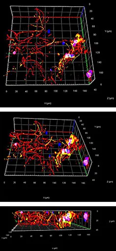
The researchers transformed skin-derived human stem cells into motor neuron progenitors—immature cells that can evolve into fully functional motor neurons. By embedding these cells into a soft, gelatin-like bioink, they utilized advanced 3D printing technology to layer the cells for optimal growth and survival. This method enhances cell survival rates and promotes nerve fiber development.
Previous methods saw limited success as nerve growth only occurred on the surface. By opting for a softer bioink conducive to internal growth, the researchers achieved a significant advancement. They further enhanced cell maturity by including mesoporous silica particles filled with growth factors, leading to enriched organoid development.
The team shared a detailed protocol for replicating these nerve tissue models, emphasizing the importance of producing a high volume of reproducible organoids. This innovative technique also opens doors to incorporating glial cells, leading to more comprehensive spinal cord models.
Key Advances in 3D Printing for Nerve Repair
The realm of 3D printing isn't just seeing innovations in ALS research. In 2018, researchers at the University of Saskatchewan made significant headway in repairing damaged peripheral nerves. Their discovery indicated that 3D printed tissue scaffolds could effectively regenerate nerves connecting the spine to the rest of the body.
Likewise, scientists from the Chinese Academy of Sciences have implemented a bioprinting technique that could potentially cure previously untreatable spinal cord injuries. By using a custom bioink loaded with neural stem cells, they successfully produced implants that restored limb movement in paralyzed rats, signaling a promising future for human applications.
Stay Connected and Informed
Join us for the upcoming Additive Manufacturing Advantage (AMAA) event on July 10th, where leaders in aerospace, space, and defense will share invaluable insights. Don’t miss out—secure your spot today!
To keep up with the latest 3D printing breakthroughs, subscribe to our newsletter and follow us on LinkedIn and YouTube for exclusive content.
Featured image showcases motor neurons generated from human stem cells.


 Brasil (PT)
 Canada (EN)
 Chile (ES)
 Česko (CS)
 대한민국 (KO)
 España (ES)
 France (FR)
 Hong Kong (EN)
 Italia (IT)
 日本 (JA)
 Magyarország (HU)
 Norge (NO)
 Polska (PL)
 Schweiz (DE)
 Singapore (EN)
 Sverige (SV)
 Suomi (FI)
 Türkiye (TR)
 الإمارات العربية المتحدة (AR)