Groundbreaking 3D Imaging Reveals Secrets of the Human Thymus: What You Need to Know!
2024-10-31
Author: Siti
Groundbreaking 3D Imaging Reveals Secrets of the Human Thymus: What You Need to Know!
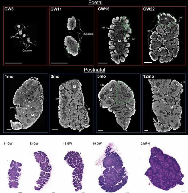
In a revolutionary advancement in medical imaging, researchers from University College London (UCL) and the Francis Crick Institute have successfully created the first-ever 3D images of a complete human thymus. Using a cutting-edge X-ray technique known as phase contrast computed tomography (PC-CT), this breakthrough offers unprecedented insights into the structure and function of this vital organ, which plays a crucial role in our immune system.
The detailed 3D images were generated from thymus samples taken from developing fetuses and infants under one year old at the European Synchrotron Radiation Facility (ESRF) in Grenoble, France. The intricate imaging revealed that structures known as Hassall's bodies take up a significant portion of the thymic medulla, leading researchers to hypothesize that these structures may influence the thymic microenvironment and immune regulation.
The thymus is instrumental in programming our immune response, particularly in the production of T cells—critical players in fighting off infections. These cells begin developing between 12-13 weeks of gestation and disseminate throughout the body, establishing a robust defense against pathogens.
Despite its importance, the thymus has often been overlooked in immunological studies. Professor Paola Bonfanti, part of the research team, emphasized, "The thymus can tell us a lot about how our immune system operates, especially regarding its transformation during early life and into adulthood." She added that methodologies like PC-CT allow researchers to maintain the organ's integrity while revealing its complex structure, crucial for understanding diseases that affect thymic architecture.
Hassall's bodies, previously thought to resemble onion layers and designated as the "graveyard" for immune cells, have been found to emerge early in organ development, filling about a quarter of the thymic medulla during peak activity in children. This suggests a significant role in shaping immune responses.
Realizing that access to high-end synchrotron facilities can be a barrier for many researchers, the team also explored a more practical method called edge-illumination. This lab-based technique mirrors the principles of synchrotron imaging while providing comparable results at a fraction of the cost and logistical complexity, turning it into a viable alternative for widespread research.
Professor Sandro Olivo highlighted the advantages of the lab-based system: "It allows us to study organ structures without damaging tissues or relying on small samples that may not accurately represent the whole organ." This opens new avenues for exploring how the thymus changes in various medical conditions, including tumor presence and age-related shrinkage.
As this cutting-edge research unfolds, it not only enhances our understanding of the thymus but could also lead to new strategies for addressing autoimmune diseases, cancers, and age-related immune deficiencies. Stay tuned for more incredible discoveries that could redefine how we approach health and medicine!


 Brasil (PT)
 Canada (EN)
 Chile (ES)
 España (ES)
 France (FR)
 Hong Kong (EN)
 Italia (IT)
 日本 (JA)
 Magyarország (HU)
 Norge (NO)
 Polska (PL)
 Schweiz (DE)
 Singapore (EN)
 Sverige (SV)
 Suomi (FI)
 Türkiye (TR)