Game-Changer in Dopamine Detection: A Revolutionary Method Combining Chemistry and Technology
2025-06-23
Author: John Tan
Innovative Breakthrough in Dopamine Detection
In an exciting advancement for neuroscience and medical diagnostics, researchers have unveiled a groundbreaking technique for detecting dopamine—a critical neurotransmitter linked to various health conditions. By leveraging the Pictet–Spengler reaction alongside surface-enhanced Raman scattering (SERS), this new approach promises to overcome long-standing hurdles in accurately measuring dopamine in biological fluids.
The Team Behind the Discovery
This innovative research hails from the prestigious Guangdong University of Technology in China, under the guidance of Kunle Li and his talented team, including Xinyu Zheng, Jiancheng Feng, Xing Feng, and Yu Zhao.
Cracking the Complex Code of Dopamine
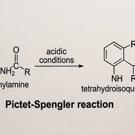
Dopamine is notoriously difficult to detect directly using SERS due to its low Raman cross-section and the intricate chemistry of physiological samples. However, the researchers have cleverly tackled these challenges by employing the Pictet–Spengler reaction, creating a chemical framework that significantly boosts both the selectivity and sensitivity of dopamine detection.
How It Works: A New Chemical Strategy
At the heart of this method lies the synthesis of aldehyde-modified layered double hydroxide. This innovative material anchors dopamine through the Pictet–Spengler reaction, which is remarkably specific to dopamine, allowing for reduced interference from other similar molecules. Additionally, silver nanoparticles functionalized with 4-mercaptophenylboronic acid serve as effective SERS probes, enabling an indirect yet highly accurate quantification of dopamine.
Outstanding Performance and Real-World Applications
The new detection method boasts exceptional analytical performance, demonstrating a linear response across a wide concentration range of 10^{-5} to 10^{10} mol·L^{-1}. Impressively, it achieves a limit of detection as low as 9.91 × 10^{-12} mol·L^{-1}, all while maintaining a strong signal-to-noise ratio of 3.
Moreover, this technique has proven effective in complex biological environments, including simulated urine and cerebrospinal fluid, highlighting its potential for clinical applications in diagnosing and monitoring neurological disorders.
A Bright Future for Neurotransmitter Research
As neuroscience continues to evolve, this novel methodology signifies a crucial step toward more effective diagnostics for mental health and neurological diseases, making it an exciting prospect for future research and clinical practices.

 Brasil (PT)
 Canada (EN)
 Chile (ES)
 Česko (CS)
 대한민국 (KO)
 España (ES)
 France (FR)
 Hong Kong (EN)
 Italia (IT)
 日本 (JA)
 Magyarország (HU)
 Norge (NO)
 Polska (PL)
 Schweiz (DE)
 Singapore (EN)
 Sverige (SV)
 Suomi (FI)
 Türkiye (TR)
 الإمارات العربية المتحدة (AR)