Breakthrough MRI Technique Unveils ‘Zombie Cells’ Behind Arthritis
2025-05-05
Author: Siti
Revolutionizing Arthritis Treatment
For anyone who's endured joint replacement surgery, the experience is often far from pleasant. Unfortunately, many osteoarthritis sufferers find themselves with limited options for relief, as current medications primarily focus on pain management, leaving the underlying progression of the disease largely unchecked.
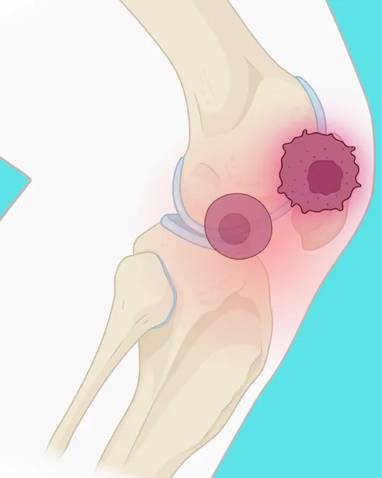
But hope is on the horizon! Enter senolytics—an innovative class of therapies that target senescent cells, often dubbed ‘zombie cells,’ that can exacerbate osteoarthritis. These dormant cells could potentially be the key to avoiding invasive surgeries altogether. While some senolytic therapies are already FDA-approved for conditions like cancer, their effectiveness for arthritis has faced a significant hurdle: researchers previously had no way to visualize these elusive cells in the body.
A Game-Changing MRI Contrast Agent
Now, a team from Stanford Medicine and Northwestern University has developed a groundbreaking MRI contrast agent that illuminates these dormant cells. This non-invasive imaging technique not only helps identify patients who could benefit from senolytic therapies but also tracks treatment progress in real-time. Their approach was recently detailed in a study published in the journal npj Imaging.
What sets this new contrast agent apart is its unique design: it’s a modified version of gadolinium, a standard MRI agent, featuring a special chemical ‘cage’ that unlocks its illuminating properties specifically in the presence of a protein called beta-galactosidase—an indicator of senescent cells. Unlike conventional gadolinium, which lights up all tissues, this innovative agent targets senescent cells exclusively.
Promising Results from Animal Trials
During initial testing on live pigs and pig knee joint specimens, the researchers observed the agent specifically lighting up the implanted senescent cells, while leaving non-senescent cells untouched. Further studies are already in the works to validate this technique in human knee specimens harvested during replacements.
The genesis of this project stemmed from a collaboration between two senior authors: Heike Daldrup-Link, MD, a radiology professor at Stanford, and Thomas Meade, PhD, a chemistry professor at Northwestern. During a chance meeting at a conference, they discovered that Meade’s existing agent could be the perfect tool for visualizing senescent cells.
Wider Applications on the Horizon
This remarkable agent doesn’t just promise advancements for arthritis; the researchers believe it can also be applied to detect and monitor senescent cells in a variety of other diseases. As aging increases the presence of senescent cells, their role in conditions ranging from cancer to Alzheimer’s and metabolic disorders becomes increasingly significant.
“Many health issues today circle back to senescence and aging,” notes Kerem Nernekli, MD, a lead author of the study. This pioneering research is supported by substantial funding from the National Institutes of Health and the NASA Ames Research Center, highlighting its potential impact on multiple fields of medicine.
A Bright Future for Arthritis Patients
With such exciting developments on the horizon, the future of arthritis treatment looks promising. The emergence of this novel MRI technique could not only enhance our understanding of joint health but also pave the way for innovative therapies that truly address the root causes of osteoarthritis. Get ready—this could be the breakthrough patients have been waiting for!


 Brasil (PT)
 Canada (EN)
 Chile (ES)
 Česko (CS)
 대한민국 (KO)
 España (ES)
 France (FR)
 Hong Kong (EN)
 Italia (IT)
 日本 (JA)
 Magyarország (HU)
 Norge (NO)
 Polska (PL)
 Schweiz (DE)
 Singapore (EN)
 Sverige (SV)
 Suomi (FI)
 Türkiye (TR)
 الإمارات العربية المتحدة (AR)