Unmasking the Silent Killer: How a Simple X-Ray Saved a Life in Aortic Dissection Case
2025-08-27
Author: Emily
The Lifesaving Importance of Quick Diagnosis
Acute aortic dissection (AD) is not just a technical term; it's a dire medical emergency where a tear in the aorta can lead to catastrophic outcomes. With death looming at a staggering rate of 1-2% per hour after symptoms begin, recognizing the signs swiftly is critical.
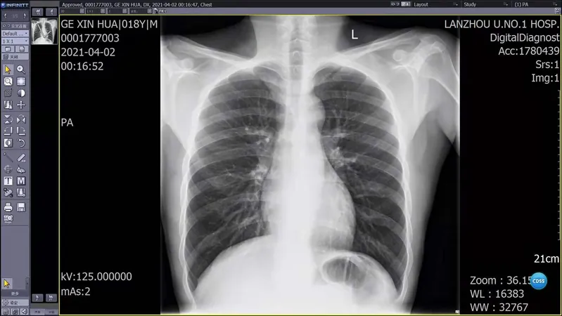
Current guidelines emphasize identifying risk factors and symptoms, including excruciating tearing pain and signs of organ distress. However, many doctors overlook the basic chest X-ray (CXR), which can reveal crucial clues such as mediastinal widening and unusual aortic contours.
A Case That Could Have Ended Tragically
Consider the case of a young man who visited a clinic after experiencing agonizing chest pain during exercise. Despite an exhaustive workup that included blood tests and a CXR, he was sent home with just pain relief, his symptoms dismissed as benign.
Three days later, as pain persisted, he returned to the emergency department during a holiday when outpatient services were shut. It was only then that a careful review of his initial CXR uncovered alarming signs—mediastinal widening and altered aortic morphology—that led doctors to perform a CT scan, ultimately diagnosing a Stanford type B aortic dissection.
Crucial Lessons Learned
This case underscores several pivotal lessons: first, that aortic dissection can manifest in unexpected ways, particularly in younger individuals who may not match the typical profile of risk factors, such as hypertension. Second, normal initial imaging findings don't rule out serious conditions. Third, in emergency medicine, reevaluating chest X-rays under optimized settings can uncover hidden dangers.
The young patient, who seemingly had no previous health issues, was quickly stabilized with beta-blockers and received urgent surgical intervention within hours.
Broadening Awareness and Training
Despite advancements in medical technology, misdiagnosis rates for aortic dissection remain alarmingly high, with studies showing that even modern healthcare systems misidentify it 14-39% of the time. This stark reality highlights the need for improved training and diagnostic protocols.
Healthcare providers should adopt standardized checklists and training to increase their awareness of atypical presentations of AD and enhance their proficiency in interpreting chest radiographs. Furthermore, emerging technologies like AI could revolutionize diagnostic accuracy by highlighting subtle radiographic signs.
Final Thoughts: A Call to Action
The journey of this young patient is a stark reminder that vigilance and meticulous attention to detail can be lifesaving. In cases presenting with chest pain, especially among younger patients, medical professionals must adhere to a 'worst-first' approach, ensuring life-threatening conditions are ruled out first.
This case serves as a rallying cry for healthcare systems worldwide to enhance diagnostic capabilities and training, ensuring that life-saving interventions are initiated when time is of the essence.


 Brasil (PT)
 Canada (EN)
 Chile (ES)
 Česko (CS)
 대한민국 (KO)
 España (ES)
 France (FR)
 Hong Kong (EN)
 Italia (IT)
 日本 (JA)
 Magyarország (HU)
 Norge (NO)
 Polska (PL)
 Schweiz (DE)
 Singapore (EN)
 Sverige (SV)
 Suomi (FI)
 Türkiye (TR)
 الإمارات العربية المتحدة (AR)