Breakthrough Imaging Technology: See Inside the Brain Like Never Before!
2025-08-10
Author: Emma
Revolutionary Brain Imaging Without Dyes!
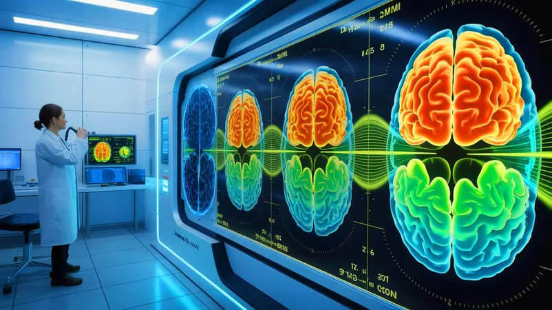
A groundbreaking advancement by MIT researchers is changing the way we explore the human brain. They’ve developed a stunning new imaging technology that allows scientists to visualize brain structures more deeply than ever, all without relying on dyes or genetic modifications!
The Power of Photoacoustic Imaging
Introducing the multiphoton photoacoustic microscope, a game-changer in neuroscience! This innovative system utilizes light to trigger sound waves, providing imaging capabilities up to five times deeper than traditional methods. It successfully penetrates dense brain tissues, enabling the study of vital molecules like NAD(P)H, which play crucial roles in cell metabolism and neuronal function.
Technical Marvel: How It Works
W. David Lee, a postdoctoral researcher involved in the design, explains that the system employs a unique three-photon excitation technique. This approach fires ultrashort bursts of light at specific wavelengths, significantly reducing scattering and optimizing deep tissue visibility. The resulting energy causes microscopic thermal expansions that produce sound waves, which are then converted into high-resolution images.
Combining Techniques for Enhanced Insights
The MIT team has ingeniously combined various advanced imaging techniques into a cohesive platform called "Multiphoton-In and Acoustic-Out." This allows for precise molecular detection while preserving tissue integrity. Notably, this system can identify crucial molecules, such as GCaMP, that track neural activity, potentially aiding in the understanding of diseases like Alzheimer’s and seizures.
Transforming Neurosurgery and Research
As researchers prepare to test this system in living animals, the excitement grows around its potential applications. The capability to map brain activity in real-time could revolutionize neurosurgery, offering surgeons invaluable insights into brain functions during procedures. Lee's previous work has shown that NAD(P)H imaging can guide wound treatment, and now its advantages could extend directly into brain surgery.
Future Challenges and Promising Directions
Although this new imaging technique promises significant advancements in neuroscience and medical technology, challenges remain. Researchers face the task of adapting this system for live subjects and ensuring its application in clinical environments. Nonetheless, the excitement surrounding this technology is palpable as scientists eagerly anticipate its potential impact on our understanding of the brain and future medical treatments.
What's Next?
Published in the journal Light: Science and Applications, this study sets the stage for further exploration of this technology. As it develops, the question arises: how will these advancements change our perception of brain function and influence the future of healthcare? Stay tuned for more incredible updates from the frontlines of science!


 Brasil (PT)
 Canada (EN)
 Chile (ES)
 Česko (CS)
 대한민국 (KO)
 España (ES)
 France (FR)
 Hong Kong (EN)
 Italia (IT)
 日本 (JA)
 Magyarország (HU)
 Norge (NO)
 Polska (PL)
 Schweiz (DE)
 Singapore (EN)
 Sverige (SV)
 Suomi (FI)
 Türkiye (TR)
 الإمارات العربية المتحدة (AR)