Unlocking the Secrets of Molecular Handedness: A Breakthrough Imaging Technique
2025-08-05
Author: Jia
Discovering Molecular Chirality
Ever wondered why spearmint and caraway seeds smell so distinct, despite having nearly identical scent molecules? Or why one version of a medicine can save lives while its mirror image could prove harmful? The answer lies in the fascinating world of chirality, essentially the "handedness" of molecules.
Revolutionary Imaging Method at ETH Zurich
A research team at ETH Zurich, helmed by Professor Romain Quidant, has made waves in the scientific community with an innovative technique that visualizes chirality in real time using just a single image. Until now, chirality measurements were largely averaged across samples, offering limited insights.
"With this groundbreaking method, we can pinpoint areas within our samples that display left-handed and right-handed chiral structures," reveals doctoral student Rebecca Büchner, who is the leading author of this promising study published in *Nature Photonics*.
Illuminating Chirality with Light
In their pioneering research, Büchner utilized specially crafted gold nanostructures, guided by lab manager Jose García-Guirado. This allowed her to anticipate the quantities of both left- and right-handed components before imaging.
The revelation of chirality came from a sophisticated imaging method akin to a specialized camera. This approach detects how samples interact with varying forms of circularly polarized light, a type of light that spirals either to the left or right.
Chiral molecules in nature respond distinctively to these light types, showing a preference for absorbing left-handed light or even altering its oscillation direction subtly.
A Game Changer for Science and Medicine
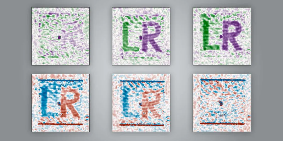
Unlike traditional imaging methods requiring two distinct measurements, Büchner's system captures both left and right circular polarizations simultaneously. By cleverly splitting light and revealing interference patterns, the method provides a clear, color-coded map that distinguishes between the left and right-handed chiral regions of samples.
This advancement enables scientists to visualize intricate structures like letters 'L' and 'R' made of nanostructures, showcasing just how detailed their observations can become.
Exploring New Frontiers in Biology and Materials Science
Jaime Ortega Arroyo, a senior scientist and co-supervisor of the project, hints at the vast potential applications: "Our technique could redefine how we measure spatial chirality, a long-standing challenge in materials science and biology."
Chirality varies within materials, and now, this method offers a way to visualize these differences instantly. In biological contexts, it could revolutionize how we assess healthy versus diseased tissues, detecting chiral differences without invasive procedures.
The implications stretch into pharmaceuticals as well; since chiral molecules are influential in drug design, this imaging could streamline the analysis and development of new therapies.
The Road Ahead: Sharpening Sensitivity
While this revolutionary imaging technique is still in the research phase, challenges remain. The current signals are moderate and vulnerable to noise.
Büchner and her team are focused on enhancing the system's sensitivity, with an eye on real-world applications. "We recognize our platform's capabilities, but collaboration with other researchers can unveil a plethora of potential use cases," she explains.
As the boundaries of chiral research are pushed, the significance of this imaging technique could reshape our understanding across multiple scientific domains.

 Brasil (PT)
 Canada (EN)
 Chile (ES)
 Česko (CS)
 대한민국 (KO)
 España (ES)
 France (FR)
 Hong Kong (EN)
 Italia (IT)
 日本 (JA)
 Magyarország (HU)
 Norge (NO)
 Polska (PL)
 Schweiz (DE)
 Singapore (EN)
 Sverige (SV)
 Suomi (FI)
 Türkiye (TR)
 الإمارات العربية المتحدة (AR)