Unlocking Heart Health: 9 Must-Know Insights from the New PET Myocardial Imaging Guidelines
2025-09-19
Author: Jia
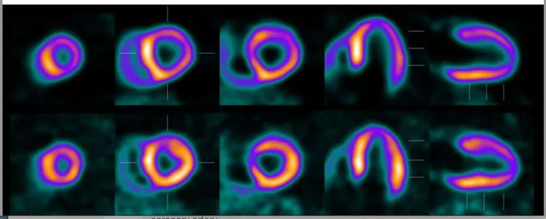
Revolutionizing Heart Imaging with PET
In the world of cardiac diagnostics, the spotlight is on 18F-flurpiridaz positron emission tomography (PET) for myocardial perfusion imaging (MPI). Today, major nuclear medicine and cardiology societies have released groundbreaking clinical guidelines aimed at refining imaging protocols and improving image interpretation.
Insights from Leading Medical Experts
Drafted by esteemed organizations, including the Society of Nuclear Medicine and Molecular Imaging (SNMMI), the European Association of Nuclear Medicine (EANM), the American College of Nuclear Medicine (ACNM), and the American Society of Nuclear Cardiology (ASNC), these guidelines are published in the Journal of Nuclear Medicine and the Journal of Nuclear Cardiology.
9 Key Takeaways
1. Enhanced Imaging Accuracy
Unlike traditional single-photon emission computed tomography (SPECT), PET imaging offers superior spatial resolution and accuracy, greatly enhancing the capacity to quantitatively evaluate myocardial blood flow (MBF).
2. Targeted Applications for Diagnosis
The guidelines recommend using 18F-flurpiridaz PET MPI for detecting flow-limiting obstructive coronary artery disease (CAD), assessing chest pain in those at intermediate to high risk of CAD, and quantifying hyperemic MBF.
3. Essential Monitoring Tools
For effective stress test monitoring, physicians should ensure the PET/CT scanner is equipped with cardiac electrocardiogram (ECG) gating, along with access to a 12-lead electrocardiograph and sphygmomanometer.
4. Optimal Injection Timing
The intravenous injection of 18F-flurpiridaz should align with the patient’s peak exercise, defined as exceeding 85% of their age-adjusted maximum heart rate. Imaging should occur shortly after injection, within 15 to 25 minutes.
5. Avoiding Conventional Modeling Pitfalls
Conventional compartment modeling cannot be employed post-exercise to calculate MBF and myocardial flow reserve (MFR) due to the already established distribution of the PET agent in the myocardium.
6. Eliminating Artifacts
To prevent misalignment artifacts, the implementation of low-dose CT for attenuation correction during rest and stress image acquisitions is highly recommended.
7. Ensuring Accurate Data Interpretation
By carefully inspecting the integrated PET and CT displays, healthcare providers can avoid misregistration that may lead to erroneous MBF calculations in critical heart regions.
8. Enhanced Diagnostic Capabilities
Preliminary research suggests that assessing stress MBF could slightly improve the diagnostic evaluation of lesions with 50-69% stenosis, potentially enhancing the detection of epicardial CAD compared to traditional MFR measurements.
9. The Future of Cardiac Imaging
As the field of cardiac imaging evolves, these new guidelines will serve as a crucial framework for clinicians, ultimately leading to better patient outcomes and revolutionizing the diagnosis of heart disease.

 Brasil (PT)
 Canada (EN)
 Chile (ES)
 Česko (CS)
 대한민국 (KO)
 España (ES)
 France (FR)
 Hong Kong (EN)
 Italia (IT)
 日本 (JA)
 Magyarország (HU)
 Norge (NO)
 Polska (PL)
 Schweiz (DE)
 Singapore (EN)
 Sverige (SV)
 Suomi (FI)
 Türkiye (TR)
 الإمارات العربية المتحدة (AR)