Shocking Case: Young Man Experiences Sudden Vision Loss Linked to Rare Leukaemia
2025-07-17
Author: Ming
Unexplained Vision Loss Brings a 33-Year-Old to the ER
In a startling medical case, a 33-year-old man rushed to the Emergency Department, gripped by panic due to a sudden, painless loss of vision in his left eye that had lasted just 1.5 hours. Previously, he had managed a history of follicular thyroid cancer, but this incident was unlike anything he had ever experienced.
What Happened Before the Crisis?
While walking, the patient described experiencing a sudden visual blackout accompanied by brief dizziness and ringing in his ears. Notably absent were common symptoms like headaches, floaters, or flashes of light.
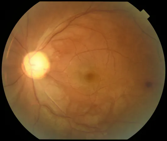
Astonishing Examination Reveals Alarming Signs
Upon examination, his left eye showed severely restricted vision—only a small part of his visual field was intact. Surprisingly, his right eye remained unaffected. Further tests revealed no foreign bodies or infections, yet a concerning relative afferent pupillary defect hinted at serious underlying issues.
Monitoring Reveals Disturbing Blood Counts
Blood tests unveiled a staggering white blood cell count of 120.55 x 10⁹/L, raising red flags for a potential severe medical condition. Initial investigations suggested myelocytes and neutrophils predominated, but other cell lines were notably diminished.
Urgent Referral and Immediate Treatment
Quickly, the patient was referred to specialized ophthalmology and hematology. Anterior chamber paracentesis was performed, and he commenced treatment with eye drops and a rigorous regimen of leucopheresis, hydroxyurea, and IV hydration.
Investigative Imaging Raises Further Alarm
An MRI of the brain indicated worrying ischemic changes in the left optic nerve and surrounding retinal tissue, hinting at a potentially severe vascular complication. A bone marrow aspiration later confirmed the diagnosis of acute leukaemia, showcasing marked myeloid hyperplasia and dysplasia.
A Grim Diagnosis: Mixed Phenotype Acute Leukaemia
The shocking diagnosis? Left eye central retinal artery occlusion (CRAO) due to hyperviscosity stemming from mixed phenotype acute leukaemia (MPAL). This rare and aggressive form of cancer could severely impact the patient’s prognosis.
Intensive Treatment and Support Are Underway
Treatment commenced with a complex chemotherapy regimen, and during hospitalization, the patient faced complications requiring multiple transfusions due to bleeding. However, there’s a glimmer of hope—slight improvements in his visual fields have been reported.
Eye-Opening Awareness for Emergency Physicians
This case emphasizes the critical need for emergency doctors to broaden their differential diagnoses when encountering sudden vision loss, particularly in younger patients. The necessity for a comprehensive hematological examination can not only identify the underlying cause but also facilitate timely, vision-preserving interventions.
A Rare Insight Into Leukaemia's Unusual Presentations
Literature suggests that while visual loss due to leukaemia is rare, it’s not unheard of. This case falls into a narrow subset of conditions already established in medical records, highlighting the urgent need for awareness and prompt action. In a world where late-stage diagnosis can mean the difference between vision and blindness, vigilance is key.


 Brasil (PT)
 Canada (EN)
 Chile (ES)
 Česko (CS)
 대한민국 (KO)
 España (ES)
 France (FR)
 Hong Kong (EN)
 Italia (IT)
 日本 (JA)
 Magyarország (HU)
 Norge (NO)
 Polska (PL)
 Schweiz (DE)
 Singapore (EN)
 Sverige (SV)
 Suomi (FI)
 Türkiye (TR)
 الإمارات العربية المتحدة (AR)