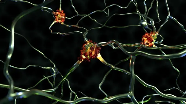
Shocking Breakthrough Reveals 'Superspreader' Fibrils in Alzheimer's Disease
2024-10-28
Author: Yu
The Battle Against Dementia Disorders
The battle against dementia disorders, especially Alzheimer's disease, continues to be one of the most daunting challenges in modern medicine. Recent research has turned the spotlight on the role of specific proteins, notably the amyloid beta protein, which tend to accumulate in the brains of affected individuals. Scientists suspect these proteins are pivotal in the onset and progression of these neurodegenerative diseases, making them a prime target for innovative therapeutic strategies.
For years, it has been known that misfolded proteins can aggregate into fibrous structures in the brain. However, a critical question about how these fibrils are formed remained unanswered — until now. A groundbreaking study led by Empa researcher Peter Nirmalraj and a team from the University of Limerick has employed advanced imaging techniques to observe this process with astonishing detail. Their findings, recently published in the journal Science Advances, put forward a startling concept: some of these slender fibrils act as "superspreaders," facilitating the spread of disease within brain tissue.
Toxic Fibrils with a Twist!
What distinguishes the so-called 'superspreader' fibrils is their remarkable properties. They exhibit heightened catalytic activity on their edges and surfaces, where new protein building blocks readily accumulate. This accumulation leads to the formation of new, extended fibrils that can propagate throughout the brain, forming harmful aggregates linked to Alzheimer's disease.
Despite knowing the chemical structure of misfolded amyloid beta protein, the formulation and characteristics of these second-generation fibrils presented a scientific enigma. Conventional methods often used for studies, like staining techniques, could distort the natural form of the proteins. Nirmalraj stated, “These interventions compromise our ability to study proteins in their true condition.”
A Cutting-Edge Approach
In this pivotal research, Empa's team utilized a state-of-the-art atomic force microscope capable of analyzing proteins in an unaltered environment — specifically, in a salt solution that closely mimics human physiological conditions. This allowed researchers to capture the fibrils, measuring less than 10 nanometers in thickness, with unprecedented precision and at room temperature.
For the first time, the team could witness the dynamics of fibril formation in real-time, documenting changes over 250 hours. Subsequent molecular modeling strengthened their findings, enabling them to categorize fibrils into distinct subpopulations based on their surface structures. “This progress marks a significant advancement in our understanding of how these proteins proliferate within the brain’s tissue in Alzheimer's patients," Nirmalraj expressed with optimism.
Implications for the Future
This research holds promise not only for deepening our understanding of the mechanics behind Alzheimer’s disease but also for informing future diagnostic approaches and methods to monitor disease progression. As the scientific community races toward identifying effective treatments, these findings may illuminate new pathways for intervention.
Funded by the Dementia Research Switzerland – Synapsis Foundation, this study represents a crucial leap forward in Alzheimer’s research, potentially paving the way for breakthrough therapies aimed at halting or even reversing the effects of this devastating condition. Stay tuned as this story develops; the implications could redefine how we approach and treat Alzheimer's disease!


 Brasil (PT)
 Canada (EN)
 Chile (ES)
 España (ES)
 France (FR)
 Hong Kong (EN)
 Italia (IT)
 日本 (JA)
 Magyarország (HU)
 Norge (NO)
 Polska (PL)
 Schweiz (DE)
 Singapore (EN)
 Sverige (SV)
 Suomi (FI)
 Türkiye (TR)