Revolutionizing Rheumatoid Arthritis Diagnosis: The Power of Thermographic Imaging!
2025-08-30
Author: Siti
Unlocking the Secrets of Inflammation with Heat Detection
In the battle against rheumatoid arthritis (RA), one critical weapon is the understanding of inflammation. Among the five classical signs of inflammation—heat, redness, pain, swelling, and loss of function—heat is a key indicator. Infrared thermography (IT), a non-invasive imaging technique, captures the infrared energy emitted by the skin, translating this into temperature readings. This has opened new doors for the detection of RA-related inflammation at the surface of joints, particularly the knees.
The Rise of Infrared Thermography in Joint Assessment
Over the last decade, studies have increasingly highlighted the effectiveness of IT, especially focused on smaller joints like hands and wrists. However, new insights are suggesting that larger joints, like the knees, deserve attention too. While previous research linked thermographic temperature increases to ultrasound-detected inflammation in smaller joints, our latest investigation dives deeper into how IT compares against ultrasound measures in assessing knee joint inflammation in RA patients.
A Study to Illuminate Knee Inflammation
This groundbreaking study recruited RA patients from a local rheumatology clinic. With informed consent and ethical approval secured, patients aged 21 to 99 had their health parameters collected, including the disease activity score and demographic data. The unique aspect? We conducted both thermographic imaging and ultrasound on the same visit, allowing us to juxtapose findings directly.
Simultaneous Imaging: A Dual Approach to Diagnosis
Each patient underwent scans on their knees using a high-performance thermal camera and a state-of-the-art ultrasound device. Both methods were executed by separate professionals blinding them to each other's findings, ensuring unbiased results. Our approach focused on meticulous accuracy in capturing joint temperatures, enabling us to assess heat patterns systematically.
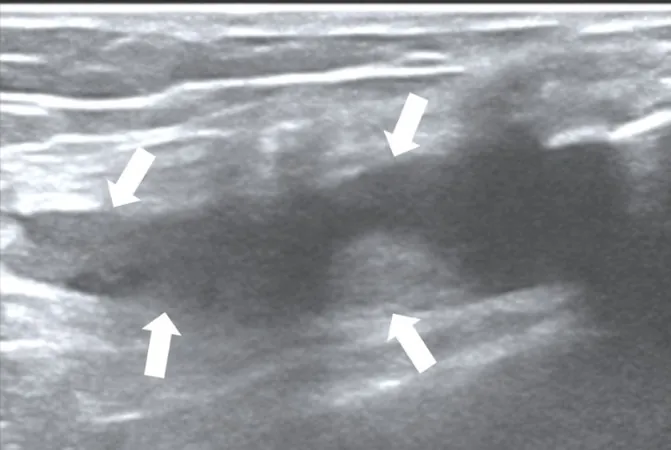
Key Findings: A New Dimension in Diagnosing RA
The analysis revealed striking correlations between thermographic parameters and ultrasound outcomes, providing a metric for identifying inflammation severity. Our results indicate that thermographic temperatures reflect joint inflammation as assessed by both gray-scale and power Doppler ultrasound, showcasing the potential of IT as an invaluable tool in clinical settings.
Reliability and Efficiency: The Thermography Advantage
While traditional imaging methods like ultrasound and MRI hold value, they can be expensive and resource-intensive. Our study found thermography to be not only cost-effective but also straightforward to use, making it an excellent option for busy clinical environments.
Recommendations for Future Research
While our findings are promising, they highlight the necessity for further exploration. Future research should aim to incorporate a control group for better comparative analysis and may expand on assessing structural joint damage alongside inflammation. Additionally, investigations into how different imaging angles at the knee affect thermography outcomes would offer deeper insights.
Conclusion: Embracing Innovation in RA Management
This study underscores the emerging role of thermography as a feasible imaging alternative for the assessment of knee joint inflammation in rheumatoid arthritis. As we continue to investigate this technology, we may revolutionize how we diagnose and monitor RA, paving the way for better patient outcomes.


 Brasil (PT)
 Canada (EN)
 Chile (ES)
 Česko (CS)
 대한민국 (KO)
 España (ES)
 France (FR)
 Hong Kong (EN)
 Italia (IT)
 日本 (JA)
 Magyarország (HU)
 Norge (NO)
 Polska (PL)
 Schweiz (DE)
 Singapore (EN)
 Sverige (SV)
 Suomi (FI)
 Türkiye (TR)
 الإمارات العربية المتحدة (AR)