Revolutionizing Lung Tumor Detection: AI Boosts Speed and Accuracy
2025-06-02
Author: Rajesh
A Surge in Lung CT Scans Worldwide
Lung disease detection is on the rise globally, with thoracic CT scans becoming a critical tool for early diagnosis of ailments like bronchial carcinoma and tumor metastases. In Germany alone, the number of lung CT scans skyrocketed from 800,000 in 2009 to a staggering 1.3 million by 2020.
The Challenge of Accurate Scan Comparisons
While these scans provide invaluable insights into treatment effects, comparing them is a laborious task fraught with potential errors. Radiologists often work under intense time constraints, complicating the evaluation of scans. However, a process known as registration—automatically aligning anatomical features across scans—can streamline this procedure.
AI to the Rescue: The SPIRABENE Project
Enter the SPIRABENE project, a groundbreaking initiative that combines the expertise of Fraunhofer MEVIS, jung diagnostics GmbH, and University Medical Center Mainz. This collaborative effort harnesses the power of artificial intelligence to enhance diagnostic accuracy and ease the workload for healthcare professionals.
"Our deep learning software can pinpoint and measure lung lesions with remarkable precision and speed, detecting even potential new lesions," states Jan Moltz, Key Research Engineer at Fraunhofer MEVIS.
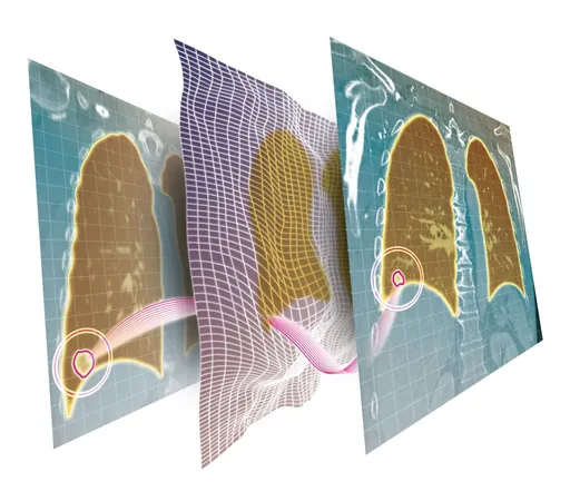
Comparing Scans with Unmatched Precision
The software compares previous CT scans with the most recent images to ensure anatomical correspondence. This process is complex, as even scans of the same patient can differ due to factors like breathing variations or weight changes.
Faster and More Efficient Detection
Thanks to deep learning technology, the automated registration process has become significantly faster and more accurate. Researchers optimized neural networks to enhance scan comparisons, and early results are promising. "AI has boosted tumor detection by 11% in follow-up images compared to traditional methods, all in under one second—just a fraction of the time required before," Moltz explains. This efficiency not only saves time but also reduces energy consumption.
Ready for Real-World Application
Developed in collaboration with UM doctors, this fully automated image processing technology has already been tested in clinical settings and is poised for real-world use. The ultimate goal is to extend AI-supported monitoring throughout the body.
A Game-Changer for Clinical Practice
Following successful trials, Moltz expresses his enthusiasm: "Creating software that directly benefits clinical practice and supports doctors is incredibly rewarding. Our tool helps identify ineffective treatments early, minimizing unnecessary side effects and costs, while improving patient recovery odds." This pioneering work stands to transform the future of lung cancer care and monitoring.


 Brasil (PT)
 Canada (EN)
 Chile (ES)
 Česko (CS)
 대한민국 (KO)
 España (ES)
 France (FR)
 Hong Kong (EN)
 Italia (IT)
 日本 (JA)
 Magyarország (HU)
 Norge (NO)
 Polska (PL)
 Schweiz (DE)
 Singapore (EN)
 Sverige (SV)
 Suomi (FI)
 Türkiye (TR)
 الإمارات العربية المتحدة (AR)