Revolutionizing Brain Imaging: How New Mathematical Models Are Fixing MRI Errors!
2025-08-08
Author: John Tan
Transforming MRI Brain Imaging with Innovative Models
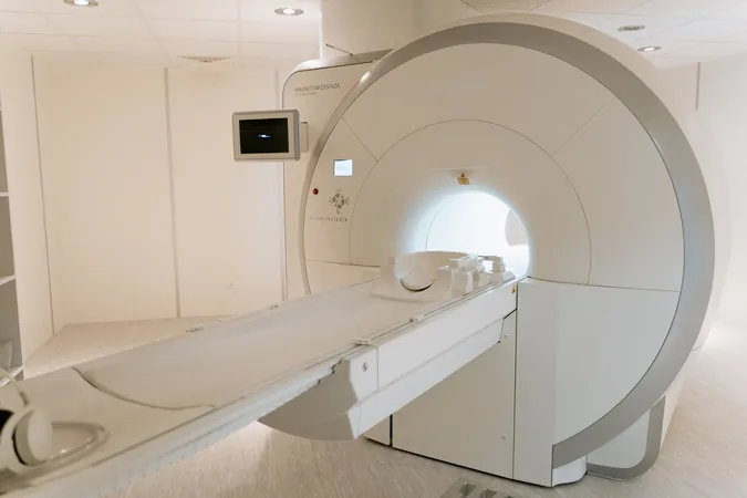
A groundbreaking team at Children's Hospital Los Angeles, led by Dr. Eamon Doyle from the Borzage Laboratory, is rewriting the rules of magnetic resonance imaging (MRI). Their cutting-edge computational models not only enhance the accuracy of cerebral blood flow imaging in both children and adults but also tackle the issue of missing data head-on.
The Challenge of Brain Imaging
When it comes to imaging the human brain, accuracy is critical, yet various factors can lead to inaccuracies. From human errors and patient movements to the complex nature of vascular structures, obtaining precise blood flow data can be a daunting task. Dr. Doyle explains, "It's pretty unlikely you'll get all four blood vessels of the brain perfectly. The technician may not notice, depending on the severity of the error."
Why Accurate Blood Flow Matters
Detecting impairments in cerebral blood flow is crucial, as these anomalies can indicate underlying brain injuries or diseases. The team's research, published in the esteemed journal Frontiers in Physiology, aims to develop mathematical models that can effectively estimate blood flow even when certain vessels are not measured accurately.
How It Works
The researchers analyzed a robust dataset of 258 phase-contrast MRIs from 108 children and 88 adults, comparing their results with a control group. This pediatric cohort included patients dealing with seizures, epilepsy, and tumors. The study meticulously examined factors like age, vessel condition, and blood flow quality.
A Game-Changer for Diagnostic Tools
Dr. Matthew Borzage, the study's corresponding author, noted the significance of these models: "You can effectively repair and utilize the data, even with a partial dataset." He further implies that while specialized equipment is usually necessary for brain imaging, these new models might enable clinicians to utilize standard 3T phase-contrast MRIs—typically used for heart imaging—to explore the intricacies of brain vessels.
Future Directions: Automation and Real-Time Analysis
Looking ahead, the team envisions automating the analysis for real-time corrections of imaging errors. Dr. Borzage emphasized, "One of the strengths of this study is that we used a varied group of kids and adults, helping us uncover overarching patterns in the population. We can produce remarkably accurate blood flow metrics to identify anomalies, enhancing diagnosis and treatment options."
The Implications Are Huge!
This innovative research marks a significant leap in medical imaging, promising to not only refine how brain conditions are diagnosed but also to improve patient outcomes across diverse demographics. As these models gain traction, they may fundamentally change the landscape of cerebral imaging, providing a more reliable tool for detecting critical brain health issues.


 Brasil (PT)
 Canada (EN)
 Chile (ES)
 Česko (CS)
 대한민국 (KO)
 España (ES)
 France (FR)
 Hong Kong (EN)
 Italia (IT)
 日本 (JA)
 Magyarország (HU)
 Norge (NO)
 Polska (PL)
 Schweiz (DE)
 Singapore (EN)
 Sverige (SV)
 Suomi (FI)
 Türkiye (TR)
 الإمارات العربية المتحدة (AR)