Revolutionizing Brain Imaging: A Breakthrough in Targeted Delivery of Agents and Drugs
2025-04-16
Author: John Tan
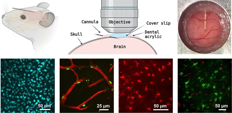
Unlocking the Secrets of the Brain with Multiphoton Microscopy
Multiphoton microscopy stands at the forefront of neuroscience, allowing researchers to dive deep into the brain's intricate activities in real time. This cutting-edge method relies heavily on the delivery of specific chemical compounds—be it imaging agents or therapeutic drugs. Unfortunately, the blood-brain barrier often thwarts these compounds from entering the brain when administered systemically.
A Game-Changer for Brain Imaging and Drug Delivery
In an exciting development, scientists at Massachusetts General Hospital have engineered a breakthrough cannula delivery system. This innovative tool enables the precise administration of imaging agents during extended in vivo imaging sessions via multiphoton microscopy. The design features a low-profile micropipette, angled as gently as 8 degrees to lie almost parallel to the brain's surface, ensuring that the optical path remains unobstructed.
Ensuring Continued Functionality and Precision
Steven Hou, the lead researcher on this project, shed light on the need for this advancement: 'Many fluorescent sensors vital for observing biological processes are not genetically encoded. This necessitates direct injection of fluorescent dyes into the brain, previously hindering long-term imaging studies. Our approach allows for a chronic, shallow-angle implantation of the cannula, facilitating the ongoing introduction of imaging agents during extensive in vivo imaging.'
Exciting Validation of the Cannula Method
To test their method's effectiveness, the research team conducted a range of experiments. They successfully delivered fluorescent cell markers to the brain while capturing dynamic images through multiphoton microscopy. In a compelling application involving mice with Alzheimer's disease, they monitored degenerating neurons using Fluoro-Jade C dye and conducted long-term imaging of oxygen levels in brain tissues with a phosphorescent sensor.
A Leap Forward in Neuroscience Research
Andy Shih, Associate Editor of Neurophotonics, lauded the technique as a major advancement for cranial imaging in mice: 'This opens new avenues for improved dye delivery and the quality of imaging data, crucial for better understanding brain functions and diseases.'
A New Era for Longitudinal Brain Studies
While not entirely noninvasive, this pioneering technique promises to elevate longitudinal studies on brain function, disease progressions, and potential remedies. It equips researchers with refined tools for exploring an expansive range of brain imaging applications. To facilitate broader adoption, the authors have generously detailed the construction and implantation process of the new cannula.


 Brasil (PT)
 Canada (EN)
 Chile (ES)
 Česko (CS)
 대한민국 (KO)
 España (ES)
 France (FR)
 Hong Kong (EN)
 Italia (IT)
 日本 (JA)
 Magyarország (HU)
 Norge (NO)
 Polska (PL)
 Schweiz (DE)
 Singapore (EN)
 Sverige (SV)
 Suomi (FI)
 Türkiye (TR)
 الإمارات العربية المتحدة (AR)