Revolutionary Thinner-Than-Eyelash Imager Set to Transform Brain Activity Monitoring
2025-05-19
Author: Arjun
A Breakthrough in Biomedical Imaging
In a groundbreaking development, researchers have unveiled an astonishingly thin and flexible imager that could revolutionize how we capture images from inside the human body. This innovative technology paves the way for early disease detection, potentially leading to faster and more effective treatment options.
As Thin as an Eyelash, Yet Powerful
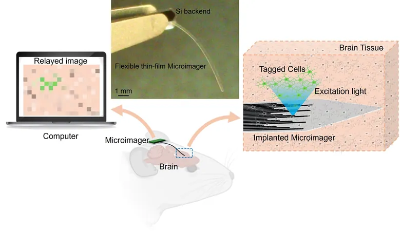
Led by Maysam Chamanzar from Carnegie Mellon University, this microimager measures a mere 7 microns thick—about a tenth of the width of a human eyelash—and 10 mm long. Its compact design allows it to navigate deep regions of the body, minimizing any risk of tissue damage, in stark contrast to the bulky and invasive endoscopes currently in use.
From Concept to Reality: Imaging the Brain
The findings, published in the journal Biomedical Optics Express, prove that this tiny device can perform structural and functional imaging within mouse brains. Remarkably, it can be tailored for varying fields of view and resolutions, opening possibilities for a multitude of medical applications.
A Game-Changer for Medical Procedures
Chamanzar envisions this microimager being implanted for short or long-term imaging, or being attached to catheters for real-time imaging of internal organs like the gastrointestinal tract or blood vessels. His team believes that integrating this technology with surgical tools could drastically enhance surgical precision and outcomes.
Innovative Use of Biocompatible Materials
The tiny imager operates on a flexible photonic platform crafted from the biocompatible polymer Parylene. This material was originally used in creating miniaturized implantable devices for focused light delivery in tissues. By leveraging Parylene waveguides, the researchers developed a sophisticated array capable of both illuminating tissue and capturing the backscattered light to generate high-resolution images.
Pioneering 3D Brain Imaging
The researchers successfully demonstrated the microimager’s capabilities by imaging fluorescent microspheres and mouse brain tissues expressing green fluorescent protein. They even captured functional neural activity, marking a significant milestone in observing neural functions in real-time.
Next Steps: A Fully Integrated Microimager
With aspirations to fully integrate light sources, image sensors, and filters into a self-contained device, the team aims to create a standalone microimager. Such an advancement could lead to vital clinical applications, such as tracking cancer cells post-surgery or monitoring disease progression after treatment.
A Vision for the Future of Neuroscience
The journey doesn’t end here. This groundbreaking work lays the foundation for correlating neural activity with specific cell types, a key desire of researchers in advancing our understanding of brain function and disease.

 Brasil (PT)
 Canada (EN)
 Chile (ES)
 Česko (CS)
 대한민국 (KO)
 España (ES)
 France (FR)
 Hong Kong (EN)
 Italia (IT)
 日本 (JA)
 Magyarország (HU)
 Norge (NO)
 Polska (PL)
 Schweiz (DE)
 Singapore (EN)
 Sverige (SV)
 Suomi (FI)
 Türkiye (TR)
 الإمارات العربية المتحدة (AR)