Revolutionary Surgical Microscope Unveils 3D Imaging Magic with 48 Tiny Cameras
2025-07-07
Author: Rajesh
A Game-Changer in Surgical Precision
For over a century, surgeons have relied on traditional stereoscopic microscopes to gain depth perception during delicate procedures. These tools mimic human sight by providing slightly different images to each eye, enabling the brain to interpret 3D structures—a vital asset when navigating fragile blood vessels or intricate brain tissues.
However, this traditional method has significant drawbacks. While it offers good depth perception, surgeons often struggle to derive precise measurements from the two-dimensional imagery presented.
The Limitations of Conventional Imaging
Accurate estimations of distances or shapes in the complex landscape of surgery—where lighting can be uneven and tools might obscure the view—remain a challenge. While preoperative 3D scans like MRIs or CTs can assist, they fail to adapt during surgery when tissues shift. Meanwhile, optical coherence tomography (OCT) delivers real-time data, but only over limited areas and in monochrome, posing interpretative hurdles.
Enter the FiLM-Scope: The Future of Surgical Tools
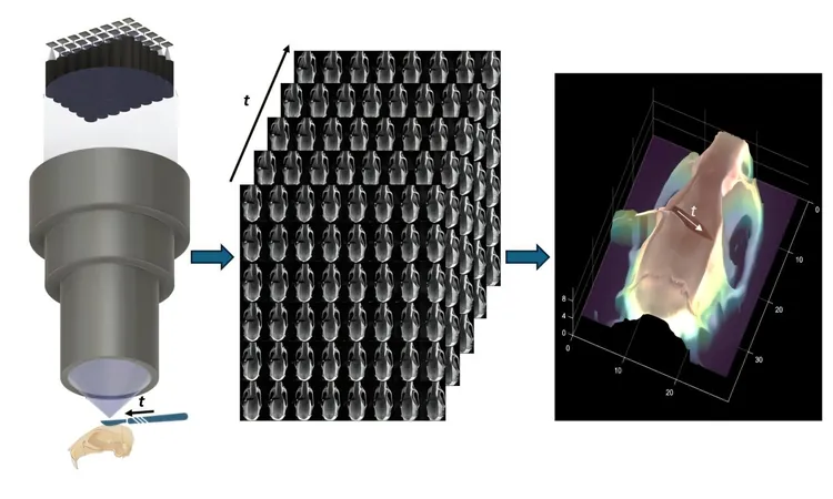
To bridge this gap, researchers have introduced a revolutionary surgical microscope: the Fourier Lightfield Multiview Stereoscope, or "FiLM-Scope." This groundbreaking device, detailed in Advanced Photonics Nexus, employs 48 mini cameras arranged in a grid—each capturing a high-resolution image (12.5 megapixels) of the surgical scene from different angles. The expansive field of view spans approximately 28 by 37 millimeters, featuring astonishing detail down to 22 microns.
Real-Time 3D Mapping at Unprecedented Speeds
With the capability to stream video at 120 frames per second, the FiLM-Scope processes these multiple perspectives through a revolutionary self-supervised algorithm that constructs a detailed 3D map of the scene in real-time. Remarkably, this algorithm functions without needing preexisting data or models, achieving surface shape reconstructions with an accuracy of 11 microns over a depth range of one centimeter.
Transforming Surgical Practices and Beyond
One of the standout features of the FiLM-Scope is its ability to allow users to digitally zoom or shift perspectives effortlessly without repositioning the microscope. This innovative tool promises to enhance the efficiency and smoothness of surgical procedures, reshaping the future of both manual and robotic microsurgery. Beyond surgery, its precise and adaptive imaging capabilities could significantly benefit fields like materials science and microfabrication, opening new realms of possibilities.


 Brasil (PT)
 Canada (EN)
 Chile (ES)
 Česko (CS)
 대한민국 (KO)
 España (ES)
 France (FR)
 Hong Kong (EN)
 Italia (IT)
 日本 (JA)
 Magyarország (HU)
 Norge (NO)
 Polska (PL)
 Schweiz (DE)
 Singapore (EN)
 Sverige (SV)
 Suomi (FI)
 Türkiye (TR)
 الإمارات العربية المتحدة (AR)