Revolutionary PET Technique Unveils Early Warning Signs of Pulmonary Arterial Hypertension
2025-01-22
Author: Ming
Introduction
In groundbreaking news, a novel imaging method known as 18F-FAPI PET has emerged as a game changer in the early detection of pulmonary arterial hypertension (PAH), a life-threatening condition that leads to dangerously high blood pressure in the pulmonary arteries. This cutting-edge technique could revolutionize the management of PAH by allowing for early intervention that can significantly improve patient outcomes.
Research Findings
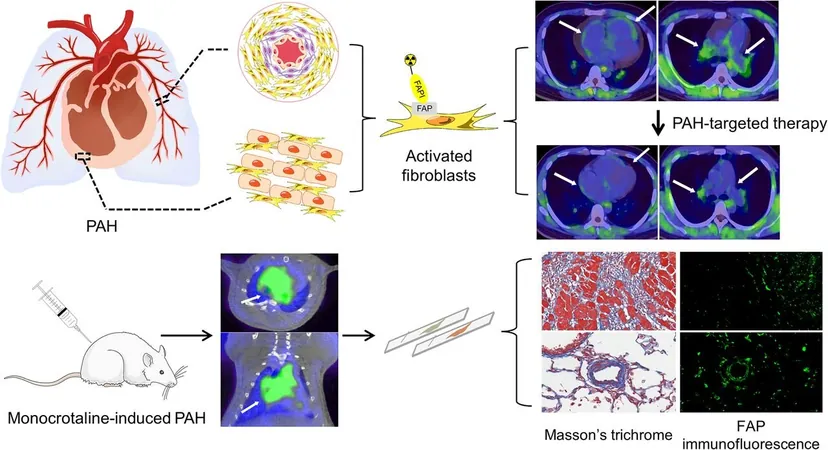
Recent research published in the Journal of Nuclear Medicine highlights the promising potential of 18F-FAPI PET to identify the initial stages of tissue remodeling—a key factor in the progression of PAH. The study emphasizes that detecting these early signs is crucial, as they often signal that irreversible structural changes are on the horizon.
Characteristics of PAH
PAH is characterized by a progressive increase in blood pressure within the arteries that supply blood from the heart to the lungs. This increase is often caused by damage or narrowing of these arteries, leading to a cascade of severe health issues. “During the remodeling process, we see significant activation of fibroblasts, which are crucial indicators of disease progression,” explains Dr. Cheng Hong, a pulmonary vascular medicine expert.
Evaluation Methods
Historically, PAH was evaluated primarily through methods such as hemodynamic measurements and echocardiography. However, the researchers behind this study aimed to explore whether imaging techniques focusing on fibroblast activation proteins could provide earlier insights into the disease’s progression.
Study Design
The study had both preclinical and clinical stages. In the preclinical phase, 15 rats were induced with PAH while a control group was established. Over the course of three weeks, significant data was collected through 18F-FAPI PET imaging and hemodynamic measurements, allowing for an evaluation of how the imaging technique correlated with the disease state.
Clinical Phase
For the clinical phase, 38 patients diagnosed with PAH underwent 18F-FAPI PET imaging, alongside right heart catheterization and echocardiography performed within a week to assess their pulmonary hemodynamic profiles. Remarkably, increased uptake of the imaging technique in the right side of the heart and pulmonary arteries correlated with their hemodynamic status and cardiac function.
Findings from Clinical Trials
Notably, three patients who received targeted PAH treatment showed a notable decrease in 18F-FAPI uptake after therapy, reflecting a positive clinical response.
Conclusion and Future Implications
“The detection of fibroblast activation provides a significant early marker for PAH progression. Our findings indicate that 18F-FAPI PET not only helps in monitoring disease status but also suggests a potential for reversibility in fibroblast activation once targeted therapies are initiated,” asserts Dr. Xinlu Wang, a nuclear medicine specialist. This innovative PET imaging approach has profound implications for the future of PAH management, highlighting the need for more personalized treatment plans. As PAH continues to pose serious health risks, advancements like 18F-FAPI PET can play an essential role in improving the prognosis for patients globally, marking an exciting new era in the field of pulmonary medicine. Stay tuned, as the implications of this research could lead to a paradigm shift in how we view and manage this daunting disease!


 Brasil (PT)
 Canada (EN)
 Chile (ES)
 Česko (CS)
 대한민국 (KO)
 España (ES)
 France (FR)
 Hong Kong (EN)
 Italia (IT)
 日本 (JA)
 Magyarország (HU)
 Norge (NO)
 Polska (PL)
 Schweiz (DE)
 Singapore (EN)
 Sverige (SV)
 Suomi (FI)
 Türkiye (TR)
 الإمارات العربية المتحدة (AR)