Revolutionary Optical Device Breaks Barriers in Super-Resolution Imaging!
2025-03-10
Author: Nur
In a groundbreaking development, researchers from the University of Science and Technology of China (USTC) have introduced a compact optical device that dramatically enhances dark-field microscopy, pushing the boundaries of super-resolution imaging beyond the traditional diffraction limit. Spearheaded by Prof. Zhang Douguo, this groundbreaking study has been published in the prestigious Proceedings of the National Academy of Sciences.
Dark-field microscopy is a well-established technique renowned for its ability to visualize unstained biological samples. By illuminating these samples at oblique angles, researchers can achieve high-contrast images of weakly scattering objects. However, conventional dark-field setups often fall short due to the diffraction limit, rendering them bulky and complicated, requiring meticulous alignment. Conversely, super-resolution imaging techniques exist, but they usually come at a high cost and involve complex operational protocols. Until now, there has been a pressing need for a more straightforward and accessible imaging solution.
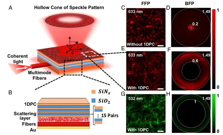
The innovative study proposes a planar photonic device that merges a scattering layer, a one-dimensional photonic crystal (1DPC), and a metallic film to generate dark-field speckle patterns. Remarkably compact, this device can seamlessly integrate into standard microscopes, effectively simplifying the overall setup and eliminating the arduous need for precise optical alignments.
The true genius of this device lies in the 1DPC, which functions as a momentum-space filter, creating hollow cones of speckle patterns. These patterns serve as an exceptional illumination source that enhances imaging quality, resulting in a 1.55-fold increase in spatial resolution compared to traditional microscopy methods.
Demonstrating the device's prowess, researchers successfully imaged a variety of samples, including polystyrene beads, nanowires, and biological specimens. Utilizing a state-of-the-art Blind-SIM reconstruction algorithm, they achieved astonishing results by resolving adjacent beads with a center-to-center distance as minuscule as 340 nanometers—exceeding the diffraction limit! The versatility of the device is further highlighted by its ability to support optical surface imaging. By tuning the incident wavelength, it can generate evanescent speckles, expanding potential applications significantly.
The experimental work used a standard upright microscope equipped with a 40x objective lens and a coherent laser source linked to multimode fibers. By dynamically vibrating these fibers, the speckle patterns could be altered, enabling the capture of multiple frames for enhanced image reconstruction. The results showcased that not only does this advanced technique elevate resolution, but it also preserves high contrast, even when imaging large fields of view.
This research marks a monumental advancement in microscopy technology. The newly designed compact planar device offers a practical, accessible, and affordable solution for super-resolution imaging. High-contrast, label-free microscopy is now on the horizon for a vast array of researchers and clinicians. By simplifying experimental setups and minimizing the need for complex alignments, this innovation could well democratize access to cutting-edge imaging techniques, paving the way for new discoveries in biological research and clinical applications.
Stay tuned for more updates as this revolutionary technology begins to transform the landscape of microscopy!

 Brasil (PT)
 Canada (EN)
 Chile (ES)
 Česko (CS)
 대한민국 (KO)
 España (ES)
 France (FR)
 Hong Kong (EN)
 Italia (IT)
 日本 (JA)
 Magyarország (HU)
 Norge (NO)
 Polska (PL)
 Schweiz (DE)
 Singapore (EN)
 Sverige (SV)
 Suomi (FI)
 Türkiye (TR)