Revolutionary Nanoprobe Transforms Inner Ear MRI Imaging
2025-09-03
Author: Mei
For years, doctors faced a daunting challenge in obtaining clear images of the inner ear, a delicate organ hidden deep within the skull's protective bone. Standard MRI techniques struggled to penetrate this area, leaving clinicians often guessing when diagnosing tumors, infections, or structural malformations.
But a groundbreaking advancement is on the horizon! A pioneering research team has unveiled a cutting-edge nanoparticle-based MRI contrast agent, designed to latch onto inner ear cells and cleverly slip past the body's natural defenses.
Huan Wang, a leading biomedical chemist at the Changchun Institute of Applied Chemistry and the study's senior author, explains, "Traditional imaging agents fail to reach this crucial tissue and are quickly eliminated from the body. This necessitates higher doses that increase the risk of hearing damage and toxicity." The culprit? A protective barrier known as the blood-labyrinth barrier (BLB), which selectively allows nutrients in while blocking most drugs and contrast agents.
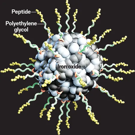
The innovation from Wang's team marks a major breakthrough. They have engineered the first guided MRI nanoprobe capable of bypassing the BLB, successfully accumulating within the inner ear. This sophisticated nanoprobe features a tiny iron oxide core measuring just 3.3 nm across for impeccable imaging capabilities, encased in a polyethylene glycol (PEG) coating to ensure prolonged circulation within the bloodstream.
But the secret weapon is a short peptide called IETP2, cleverly designed to bind with LRP1, a receptor that thrives in the BLB. This enables the nanoprobe to hitch a ride across the barrier, effectively delivering it where it's needed most.
In animal trials, the nanoprobes demonstrated remarkable efficacy, efficiently crossing the BLB and persisting in circulation long enough to target the cochlea. With MRI signals up to 84% stronger than those obtained from standard controls, this development promises clearer diagnostic insights without risking damage to the surrounding organs.
Carmen Burtea, a biomedical imaging researcher at Université de Mons, highlights the untapped potential of this nanoprobe: "The untargeted version has already shown enhanced contrast at lower doses than traditional gadolinium agents. With further tests, it could revolutionize the way we diagnose fluid and structural changes within the inner ear."
However, Wang acknowledges that translating this technology to clinical settings will necessitate rigorous toxicology assessments and larger animal studies. Experts like Johannes Gerb from Ludwig Maximilian University Munich note the probe's potential as a viable alternative to gadolinium if it proves safe and effective in humans, potentially transforming inner ear diagnostics forever!


 Brasil (PT)
 Canada (EN)
 Chile (ES)
 Česko (CS)
 대한민국 (KO)
 España (ES)
 France (FR)
 Hong Kong (EN)
 Italia (IT)
 日本 (JA)
 Magyarország (HU)
 Norge (NO)
 Polska (PL)
 Schweiz (DE)
 Singapore (EN)
 Sverige (SV)
 Suomi (FI)
 Türkiye (TR)
 الإمارات العربية المتحدة (AR)