Revolutionary MRI Technology Sets New Standard for Glioblastoma Treatment
2024-10-01
Revolutionary MRI Technology Sets New Standard for Glioblastoma Treatment
In an exciting breakthrough for cancer treatment, a recent study highlights how cutting-edge MRI technology can transform the management of glioblastoma, one of the most aggressive forms of brain cancer. Conducted by researchers at the Sylvester Comprehensive Cancer Center, part of the University of Miami Miller School of Medicine, this innovative study marks the first to analyze tumor changes in real-time for patients undergoing MRI-guided radiation therapy.
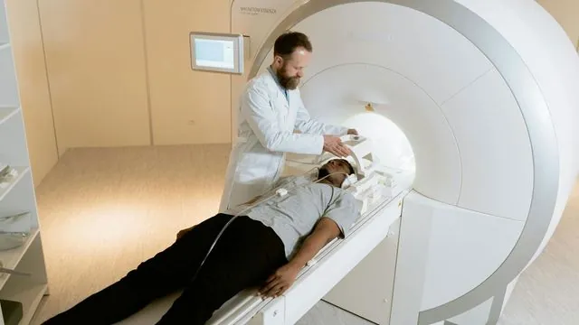
Utilizing a method known as MRI-linear accelerator (MRI-linac), this approach combines daily imaging with radiation treatment, allowing physicians to monitor tumors with unprecedented accuracy. Sylvester is recognized as a pioneer in employing this technology for glioblastoma patients and is among the few cancer centers globally to offer such advanced treatment options.
The findings, published on September 30 in the International Journal of Radiation Oncology and presented at the American Society for Radiation Oncology (ASTRO) meeting, reveal the potential of daily MRI scans to serve as an early warning system for tumor growth. This capability could fundamentally change how oncologists adapt treatments during radiation therapy, ensuring that patients receive the best possible care in real time.
Led by Dr. Eric A. Mellon, a radiation oncologist and co-leader of Sylvester’s Neurologic Cancer Site Disease Group, the study closely monitored 36 glioblastoma patients over a six-week period of daily radiation and MRI scans. By comparing these daily images to the standard imaging procedures—conducted before and after treatment—the researchers assessed the tumor size and growth patterns effectively.
Remarkably, for 74% of participants, results from the MRI-linac aligned with the traditional contrast MRI scans, confirming whether tumors grew, shrank, or remained stable. However, there were discrepancies in 26% of cases, where the real-time imaging indicated growth, but the conventional imaging reflected shrinkage. Interestingly, despite not matching all the expected outcomes, the MRI-linac successfully identified all instances of genuine tumor growth, suggesting its valuable role in active cancer treatment.
This innovative technology could also provide benefits beyond monitoring tumor growth. In cases where tumors shrink, radiation can be precisely targeted to minimize harm to surrounding healthy tissue. Additionally, as many glioblastoma patients undergo surgery prior to radiation, tracking changes in the surgical cavity during recovery can inform treatment adjustments, ultimately protecting healthy brain areas from radiation exposure.
Looking ahead, Dr. Mellon and his team plan to conduct further studies to further explore the capabilities of MRI-linac in guiding glioblastoma treatment. They hope to expand its application to other brain cancers, aiming to refine strategies that could significantly improve patient outcomes against this notoriously aggressive cancer.
In a broader context, these advancements in imaging technology signify a turning point in the battle against glioblastoma. With glioblastoma often leading to dire prognoses, this innovative approach could revolutionize the way oncologists tailor treatment to individual patients, maximizing efficacy while minimizing risks.
As we continue to witness significant strides in cancer treatment technologies, it becomes increasingly evident that the integration of real-time imaging with traditional therapies may very well shape the future of oncology, offering renewed hope to patients and their families faced with the daunting realities of brain cancer.

 Brasil (PT)
 Canada (EN)
 Chile (ES)
 España (ES)
 France (FR)
 Hong Kong (EN)
 Italia (IT)
 日本 (JA)
 Magyarország (HU)
 Norge (NO)
 Polska (PL)
 Schweiz (DE)
 Singapore (EN)
 Sverige (SV)
 Suomi (FI)
 Türkiye (TR)