Revolutionary MRI Technology Set to Transform Aortic Stenosis Diagnosis
2025-05-09
Author: Li
In a groundbreaking advancement, researchers have unveiled a state-of-the-art MRI technique that could redefine how aortic stenosis, a prevalent and potentially life-threatening heart condition, is diagnosed. This condition impacts an estimated 300,000 people in the UK and affects around 5% of individuals aged 65 and older in the US, with its prevalence rising as people age.
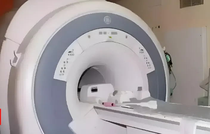
Conducted by an innovative team from the University of Sheffield in collaboration with the University of East Anglia, a recent study has demonstrated that a four-dimensional flow (4D flow) MRI can diagnose aortic stenosis with unprecedented accuracy compared to traditional ultrasound methods.
This cutting-edge technology significantly enhances the ability of doctors to determine the urgency of surgical intervention for patients. Published in the journal Open Heart, the study titled "Four-dimensional flow provides incremental diagnostic value over echocardiography in aortic stenosis" highlights the findings from examinations of 30 patients diagnosed with the condition.
Researchers employed both conventional ultrasound scans (echocardiography) and the novel 4D flow MRI technique, comparing the efficacy of each in identifying patients needing timely heart valve surgery. Validation over eight months showcased that the 4D flow MRI provided superior measurements of blood flow through the heart valves, proving to be more reliable than standard echocardiography.
Professor Andy Swift from the University of Sheffield's School of Medicine and Population Health emphasized the significant potential of this technology, stating, "4D flow scanning not only enhances our diagnostic accuracy but may lead to earlier and more precise interventions for those suffering from aortic stenosis." He also serves as an Honorary Consultant Radiologist at Sheffield Teaching Hospitals Foundation Trust.
Lead researcher Dr. Pankaj Garg, a consultant cardiologist at the Norfolk and Norwich University Hospital, expressed enthusiasm for this advancement, remarking, "Aortic stenosis is a common yet dangerous heart condition. Our innovative 4D flow MRI allows us to visualize blood flow in three dimensions over time—capturing the fourth dimension. Our goal was to determine if it could outshine traditional ultrasound in providing diagnosis accuracy."
As the medical community braces for the potential impacts of this groundbreaking technology, patients suffering from aortic stenosis may soon experience enhanced diagnostic processes, leading to better treatment outcomes and improved quality of life.


 Brasil (PT)
 Canada (EN)
 Chile (ES)
 Česko (CS)
 대한민국 (KO)
 España (ES)
 France (FR)
 Hong Kong (EN)
 Italia (IT)
 日本 (JA)
 Magyarország (HU)
 Norge (NO)
 Polska (PL)
 Schweiz (DE)
 Singapore (EN)
 Sverige (SV)
 Suomi (FI)
 Türkiye (TR)
 الإمارات العربية المتحدة (AR)