Revolutionary Morphing Brain Sensor Paves the Way for Enhanced Neurostimulation
2024-09-28
Author: Arjun
In an exciting development for neuroscience, researchers have unveiled a groundbreaking morphing brain sensor designed to enhance transcranial focused ultrasound (TFUS) capabilities.
This non-invasive technique utilizes high-frequency sound waves to stimulate targeted areas of the brain, offering promising treatment options for numerous neurological disorders, including drug-resistant epilepsy and conditions characterized by recurrent tremors.
Bridging the Gap in Brain Signal Measurement
According to Donghee Son, the study's supervising author, previous brain sensors used for direct contact with the brain faced significant challenges in accurately capturing neural signals due to their inability to conform tightly to the intricate folds of the brain.
Earlier efforts, such as those by Professors John A. Rogers and Dae-Hyeong Kim, led to some improvements, but limitations remained—particularly in areas of severe curvature, where these sensors struggled to maintain secure adhesion.
The inadequacies of existing sensors in preventing slippage due to micro-movements or fluctuations in cerebral spinal fluid have hampered their medical application. To overcome these barriers, Son and his colleagues set out to create a sensor that conforms to even the most complex brain surfaces, allowing for reliable and extended measurement periods of brain activity.
A New Chapter in Neurostimulation
The newly developed sensor, termed ECoG (electrocorticogram), showcases exceptional adherence to brain tissue without forming voids, drastically reducing interference from external mechanical movements.
This feature is crucial in optimizing the benefits of low-intensity focused ultrasound (LIFU)—a key component in treating epilepsy—where therapy must be personalized for each patient.
Son emphasized the importance of this sensor in facilitating real-time monitoring of brain waves while simultaneously delivering targeted ultrasound stimulation, enabling a tailored approach to treatment.
The noise reduction achieved through the new technology may finally bridge the gap towards developing personalized treatment protocols that cater to the individual needs of patients.
Innovative Design and Future Applications
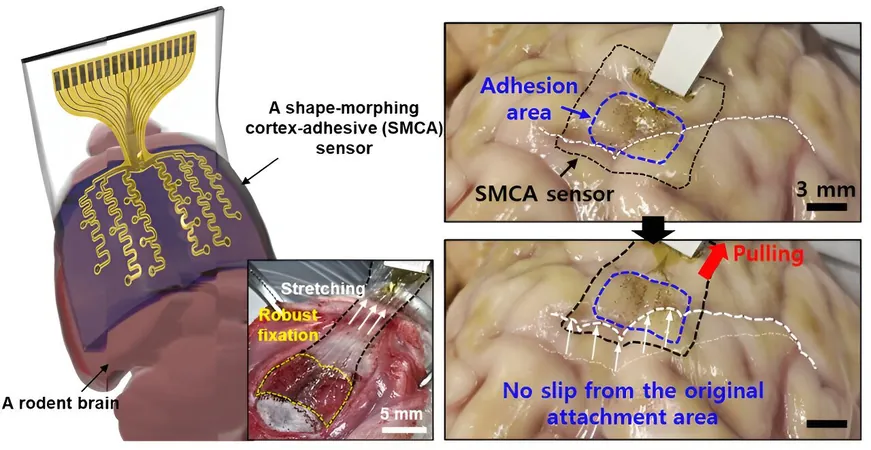
The ECoG sensor consists of a three-layer structure: a hydrogel-based layer for strong bonding with brain tissue, a self-healing polymer-based layer that can change shape to match the brain’s contours, and an ultrathin stretchable layer outfitted with gold electrodes.
This innovative incorporation allows the sensor to undergo a gelation process upon attachment to the brain, providing a secure and adaptable fit.
The sensor has shown promising results in initial tests conducted on awake rodents, demonstrating its capability to accurately measure brain waves and control seizure activities.
The research team is enthusiastic about expanding the design to create a high-density array of sensors that could significantly enhance the mapping of brain signals.
A Future of Enhanced Neurological Treatments
Future versions of this sensor aim to include a higher number of electrode channels and develop minimally invasive implantation techniques, potentially revolutionizing how neurological disorders are diagnosed and treated.
The overarching goal is to advance clinical research, bringing forth new avenues for non-invasive therapies for epilepsy and other brain disorders.
As the research progresses, the sensor could lay the foundation for more effective prosthetic technologies, ultimately transforming patient care and treatment outcomes in neurology.
Don’t Miss Out on the Future of Neuroscience!
This research highlights the immense potential of innovation in medical science, promising a new dawn for brain-related therapies that could change countless lives across the globe.
Stay tuned as we continue to uncover the advancements that hold the key to battling neurological disorders like never before!

 Brasil (PT)
 Canada (EN)
 Chile (ES)
 Česko (CS)
 대한민국 (KO)
 España (ES)
 France (FR)
 Hong Kong (EN)
 Italia (IT)
 日本 (JA)
 Magyarország (HU)
 Norge (NO)
 Polska (PL)
 Schweiz (DE)
 Singapore (EN)
 Sverige (SV)
 Suomi (FI)
 Türkiye (TR)
 الإمارات العربية المتحدة (AR)