Revolutionary Mini Imager: The Future of Brain Scans is Here!
2025-06-03
Author: Daniel
A Breakthrough in Medical Imaging
In a groundbreaking development, researchers from Carnegie Mellon University have unveiled a cutting-edge, ultra-thin imaging device that could transform how we visualize internal tissues, particularly in the brain. This remarkable microimager, thinner than a human eyelash, heralds a new era for minimally invasive diagnostics.
The Impressive Specs of the Microimager
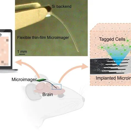
Measuring just 7 microns thick and 10 millimeters long, this innovative device offers unmatched flexibility for accessing delicate areas deep within the body. Its diminutive size means it can navigate tight spaces without inflicting damage on surrounding tissues, a significant advance over traditional endoscopic tools.
Revolutionizing Endoscopic Technology
Professor Maysam Chamanzar, the leader of this pioneering research team, emphasized that unlike conventional endoscopes—which are often bulky and cumbersome—this microimager is designed for seamless integration into minimally invasive procedures. This breakthrough means that sections of the body previously deemed unreachable might soon be easily accessed.
A Peek Inside the Brain
In laboratory trials, the team successfully employed the imager to visualize live neural activity and brain structures in mouse models. Utilizing a biocompatible and transparent polymer called Parylene, they crafted a flexible photonic system capable of guiding light in multiple directions. This ability allows the device to both illuminate tissues and capture essential data from the surroundings.
Innovative Design with Micromirrors
Each waveguide in this advanced device is equipped with micromirrors at each end. While some waveguides project light onto biological tissues, others collect the reflected light, sending it to an image sensor. This unique structure means that each waveguide acts as a pixel, creating a detailed image of what’s happening under the surface.
Promising Results in Neuroscience
The researchers demonstrated the device's capabilities by imaging fluorescent beads in different mediums to confirm its 3D localization abilities. They captured images from mouse brain tissue expressing green fluorescent protein and showcased its functionality by using calcium indicators to observe neural activity.
Validating Accuracy Against Electrophysiology
To reinforce the credibility of their imaging technique, Dr. Vishal Jain and his team compared the new data with traditional electrophysiological recordings. The striking alignment of results provided strong validation for their innovative imaging approach.
Aiming for a Comprehensive Neuroscience Tool
Professor Chamanzar outlined the long-term vision of the project: mapping neural activity alongside gene expression in specific cell types—a lofty yet vital goal in systems neuroscience.
The Future of Medical Diagnostics
Future iterations of the microimager aim to include image sensors, filters, and light sources, creating a comprehensive platform for in vivo applications. This technology could revolutionize how we detect residual cancer cells after surgery and monitor disease progression.
Unlocking New Diagnostic Possibilities
"This microimager opens the door to countless opportunities in diagnostics and real-time surgical guidance," said Chamanzar. With further refinements, this tiny device could become an indispensable asset in the field of minimally invasive medicine.


 Brasil (PT)
 Canada (EN)
 Chile (ES)
 Česko (CS)
 대한민국 (KO)
 España (ES)
 France (FR)
 Hong Kong (EN)
 Italia (IT)
 日本 (JA)
 Magyarország (HU)
 Norge (NO)
 Polska (PL)
 Schweiz (DE)
 Singapore (EN)
 Sverige (SV)
 Suomi (FI)
 Türkiye (TR)
 الإمارات العربية المتحدة (AR)