Revolutionary Microscope Tech Unveils the Hidden Depths of Brain Tissue
2025-08-24
Author: Yu
Groundbreaking Technology Developed at MIT
MIT researchers have just unveiled an innovative microscope technology that dives deeper into brain tissue than ever before, revealing the intricate details at a single-cell level. By utilizing a sophisticated blend of light and sound, this cutting-edge system generates high-resolution images without the need for external labels, marking a significant leap in biomedical imaging.
How It Works: A Two-Stage Process
The new method, dubbed "Multiphoton-In and Acoustic-Out," operates through a unique two-step process. It begins with a powerful burst of long-wavelength light that penetrates deep into brain tissue, employing a technique known as "three-photon light" to excite specific molecules within individual cells.
In the next stage, instead of relying on faint fluorescent light emissions, this innovative approach captures sound. The absorption of light energy causes thermal expansion in the cell, producing sound waves that travel through the surrounding tissue. A sensitive ultrasound microphone then detects these sound waves, transforming them into detailed images.
A Major Milestone in Imaging Technology
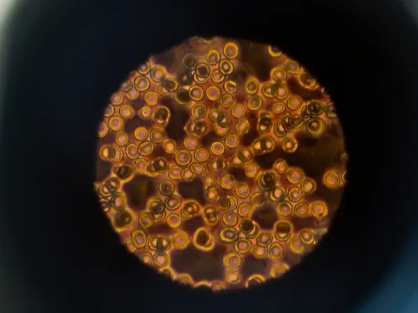
In an impressive demonstration of its capabilities, the research team successfully imaged NAD(P)H—a key molecule tied to cellular metabolism and neuron activity—through a 1.1 millimeter-thick human cerebral organoid. This achievement is noteworthy, boasting a depth of imaging five times greater than current label-free microscopy technologies.
Potential Clinical Applications
One of the most exciting aspects of this new technology is its potential for real-world applications. Since it requires no additional chemicals or genetic modifications, the researchers envision its usage in clinical settings, particularly for detecting biomarkers that indicate diseases like Alzheimer's during brain surgeries.
While initial tests have only taken place in controlled environments, the team is eager to move forward and demonstrate this groundbreaking technology on living animals, paving the way for its future use in medical diagnostics.


 Brasil (PT)
 Canada (EN)
 Chile (ES)
 Česko (CS)
 대한민국 (KO)
 España (ES)
 France (FR)
 Hong Kong (EN)
 Italia (IT)
 日本 (JA)
 Magyarország (HU)
 Norge (NO)
 Polska (PL)
 Schweiz (DE)
 Singapore (EN)
 Sverige (SV)
 Suomi (FI)
 Türkiye (TR)
 الإمارات العربية المتحدة (AR)