Revolutionary Microscope Captures Live 3D Biological Images at Lightning Speed!
2025-08-15
Author: Jia
The Future of Astrobiology is Here
As we explore distant worlds with more sophisticated astrobiology probes, the need for in-situ analysis without human assistance becomes paramount. This technology is set to transform how we study alien ecologies when humans eventually visit these far-off places.
High-Speed 3D Imaging Breakthrough
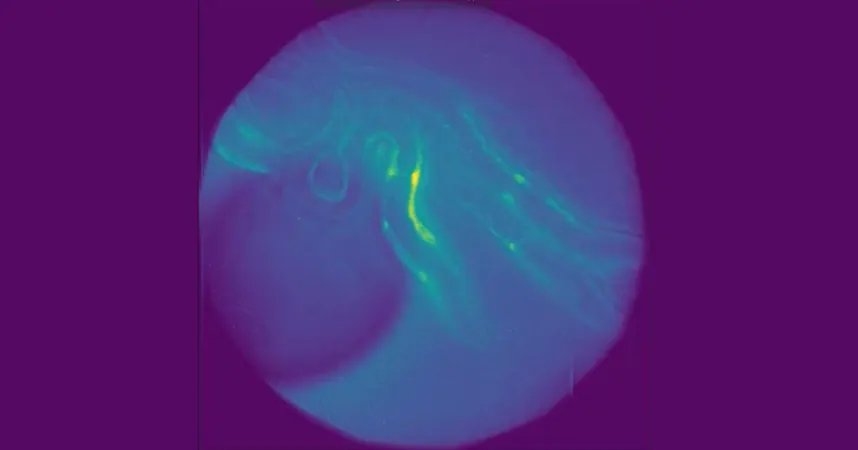
A groundbreaking microscope, capable of capturing detailed 3D images of entire small organisms in real-time, has been unveiled, promising to redefine what we know about developmental biology and neuroscience. This innovation could unlock new paths in research!
Traditional microscopes struggle with speed and depth adjustment, often resulting in distorted or incomplete images of fast-moving biological processes. Eduardo Hirata Miyasaki, who developed this technology at the University of California Santa Cruz, stated, "Our new system enhances the multifocus microscopy (MFM) technique with a 25-camera array to capture living organisms without disruption."
Meet the M25: A Microscope Like No Other
Dubbed the M25, this state-of-the-art microscope blends cutting-edge diffractive optics with 25 miniature cameras that can simultaneously observe multiple depths. This enables researchers to capture stunning live 3D volumes at speeds exceeding 100 volumes per second!
"The M25 is particularly adept at imaging C. elegans worms, often used as model organisms in various studies. Unlike previous methods that could only visualize sections of the worm, this innovative technology allows for comprehensive observation, facilitating research on nervous systems and the effects of genetic mutations or treatments," said Miyasaki.
The Magic of Multi-Plane Light Control
Central to this microscope’s power is its use of diffractive optical elements, enabling efficient light manipulation. These microstructures offer superior performance compared to traditional optics, leading to optimized imaging without the bulk of standard components.
By incorporating custom-designed gratings to manage light dispersion effectively, the M25 achieves high-resolution images while remaining compact and scalable.
Imaging Without Borders
The M25 microscope is designed for both fluorescence and label-free imaging, making it ideal for observing delicate biological systems without the need for dyes or invasive markers. This capability is especially vital in fields like embryology, where maintaining natural conditions is crucial.
To ensure the technology works flawlessly, researchers validated the microscope with prototype tests, successfully imaging multiple focal planes and live specimens, showcasing the ability to capture dynamic 3D images without distortion.
Preparing for Tomorrow's Discoveries
This system can easily integrate into existing commercial microscopes without the need for specialized hardware, making it accessible for widespread use. Looking ahead, the researchers aim to enhance this system’s capabilities further by employing machine learning to analyze imaging data for disease detection and cell behavior tracking.
The M25 represents not just a leap in microscopy but a major stride in our quest to understand life, both on Earth and beyond.
Join the Revolution in Biological Imaging!
Stay tuned as this cutting-edge technology continues to evolve, paving the way for the next frontier in biological research and astrobiology! The implications are staggering, and the journey has just begun!


 Brasil (PT)
 Canada (EN)
 Chile (ES)
 Česko (CS)
 대한민국 (KO)
 España (ES)
 France (FR)
 Hong Kong (EN)
 Italia (IT)
 日本 (JA)
 Magyarország (HU)
 Norge (NO)
 Polska (PL)
 Schweiz (DE)
 Singapore (EN)
 Sverige (SV)
 Suomi (FI)
 Türkiye (TR)
 الإمارات العربية المتحدة (AR)