Revolutionary Microfluidics Platform Unveils Secrets of Tick-Borne Babesiosis
2025-08-22
Author: Daniel
Babesiosis is not your average infectious disease; it mirrors malaria in symptoms and spreads like Lyme disease. Once a rarity in the United States, this ailment is now surging, especially across the Northeast, Mid-Atlantic, and Upper Midwest regions.
In a major breakthrough, researchers at Carnegie Mellon University have engineered an innovative microfluidics platform designed to closely monitor Babesiosis infections within red blood cells, providing critical insights into this potentially deadly disease.
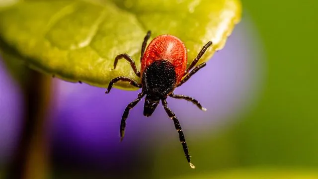
Caused by the parasite Babesia microti, Babesiosis is transmitted to humans through tick bites after these insects feast on infected rodents. Once in the bloodstream, the parasite wreaks havoc by invading and damaging red blood cells, which is especially dangerous for older adults and those with weakened immune systems.
Timely and precise diagnosis is vital, as symptoms can be vague and flu-like, making it difficult to identify without proper testing.
The Science Behind the Breakthrough
In an article published in Advanced Science, lead researcher Tagbo Niepa and his team introduce a microfluidic system that simulates the natural environment of Babesia microti, enabling more effective study outside of traditional animal models. "We wanted to create an environment where the parasite can thrive and be studied in vitro," states Niepa, who leads the Niepa μBiointerface Lab.
Microorganisms often behave differently in lab settings compared to their natural habitats, posing significant challenges for researchers. To combat this, the team developed a platform that maintains the essential microenvironment for the parasite, allowing real-time observation of its life cycle.
Innovative Design for Real-Time Observation
The microfluidics platform features a meticulously crafted channel that accommodates a single layer of red blood cells, enabling scientists to observe the infection dynamics of Babesia in real time. Its open design facilitates better physical and optical access to samples, which is a game changer for studying individual cells.
Researchers have marked the Babesia microti with fluorescent stains, making them easily distinguishable during confocal microscopy imaging. This advancement not only accelerates parasite identification but also allows for the measurement of its density in blood and the assessment of damage inflicted on red blood cells.
The Road Ahead: Integrating AI for Enhanced Detection
Niepa is now focusing on enhancing the platform's capabilities by integrating automation, imaging technologies, and AI-driven analysis. Current hospital methods to detect Babesia microti in blood smears take about five minutes with a standard microscope, but Niepa envisions a future where advanced imaging techniques, powered by AI, can expedite detection and provide invaluable data on infection levels.
"There's a lot more characterization to perform," Niepa explains. Notably, while Babesia microti affects rodents and humans, it does not infect deer, raising intriguing biological questions that could be more easily explored using this innovative in vitro platform.
Extending Sample Viability and Future Enhancements
While traditional blood samples decay over time, the system developed by Niepa and collaborator Chao Li extends the viability of samples up to 72 hours. Future improvements aim to push this boundary even further, positioning the platform as a pioneering tool for studying Babesia microti and potentially transforming the landscape of infectious disease research.

 Brasil (PT)
 Canada (EN)
 Chile (ES)
 Česko (CS)
 대한민국 (KO)
 España (ES)
 France (FR)
 Hong Kong (EN)
 Italia (IT)
 日本 (JA)
 Magyarország (HU)
 Norge (NO)
 Polska (PL)
 Schweiz (DE)
 Singapore (EN)
 Sverige (SV)
 Suomi (FI)
 Türkiye (TR)
 الإمارات العربية المتحدة (AR)