Revolutionary Imaging Technology Offers Unprecedented Access to Living Brain Cells
2025-08-22
Author: Wei Ling
Unlocking the Mysteries of the Brain
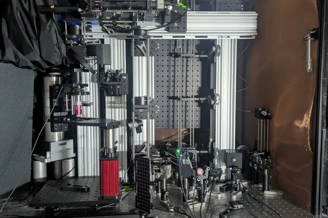
For decades, researchers have been on a quest to unveil the secrets of brain activity, diving deeper with each advancement in microscopy. Now, a groundbreaking study by a team from MIT has developed a cutting-edge microscope that can penetrate deep into brain tissues, revealing the intricate molecular activity of individual cells using sound.
A Leap Forward in Neuroscience
Leading the charge, neuroscientist Mriganka Sur, along with mechanical engineering professor Peter So and principal research scientist Brian Anthony, have ushered in a new era of imaging technology. Sur, a prominent figure at MIT’s Picower Institute for Learning and Memory, described their work as a remarkable leap in imaging capabilities, enabling unprecedented single-cell resolution.
Deep Insights into Cellular Function
In their study published in *Light: Science and Applications*, the team successfully detected NAD(P)H, a crucial molecule associated with cellular metabolism and neuronal activity, at depths more than five times greater than previous technologies. Their innovative microscope achieved this feat by utilizing a technique that allowed them to visualize through samples such as 1.1-millimeter thick cerebral organoids and mouse brain slices.
Innovative Techniques Combined
Co-lead author W. David Lee shared that while their current samples displayed impressive results, the potential for even greater depths exists. Utilizing a distinct ‘three-photon’ excitation method, the microscope focuses short bursts of light to penetrate deeper with reduced scattering. This produces localized thermal expansions within cells that generate sound waves, which the apparatus then transforms into high-resolution images.
The Future of Brain Imaging
The integration of techniques forms what they call the 'Multiphoton-In and Acoustic-Out' imaging platform. This method not only provides intricate cellular visuals through simultaneous third-harmonic generation imaging but also opens doors for detecting various molecules, including indicators of neural electrical activity.
Pathways to Clinical Applications
Looking ahead, the team aims to translate these findings into real-world scenarios. Their imaging method is notably label-free, making it ideal for human application, such as during brain surgeries. Lee’s previous ventures in NAD(P)H imaging show promise in medical settings like wound care, illustrating the molecule's variations in conditions like Alzheimer’s disease and seizures.
Next Steps in Research
As they plan to move beyond laboratory samples to live animal testing, the team anticipates achieving imaging depths of up to 2 millimeters in living brains. With confidence in their methodology, the researchers are poised to explore uncharted territories in neuroscience.
A Bright Horizon for Neuroscience
This innovative advancement not only promises to enhance our understanding of the brain’s complexities but also presents potential breakthroughs in the diagnosis and treatment of neurological disorders. With support from various research funding sources, this pioneering work will undoubtedly pave the way for future developments in brain imaging.

 Brasil (PT)
 Canada (EN)
 Chile (ES)
 Česko (CS)
 대한민국 (KO)
 España (ES)
 France (FR)
 Hong Kong (EN)
 Italia (IT)
 日本 (JA)
 Magyarország (HU)
 Norge (NO)
 Polska (PL)
 Schweiz (DE)
 Singapore (EN)
 Sverige (SV)
 Suomi (FI)
 Türkiye (TR)
 الإمارات العربية المتحدة (AR)