Revolutionary Imaging Technique Unveils Brain Changes Linked to Learning and Memory!
2025-03-31
Author: Yu
Understanding Neuronal Communication
Scientists have long grappled with understanding how neurons communicate through minuscule junctions known as synapses. This rapid signaling—taking mere milliseconds—plays a crucial role in processes like learning and memory. However, pinpointing the specific locations within the brain where synaptic changes occur during learning has remained a significant challenge.
The Breakthrough: DELTA
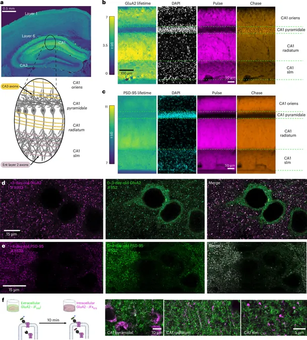
Recent breakthroughs from researchers at Janelia have paved the way for a groundbreaking imaging technique dubbed DELTA. Spearheaded by the collaborative efforts of the Spruston and Lavis labs, along with contributions from former senior researcher Karel Svoboda, this innovative method has transformed our ability to visualize the dynamic changes in synaptic connections across the entire brain.
Unprecedented Insights
Published in the prestigious journal *Nature Neuroscience*, DELTA offers an unprecedented view of how individual synaptic proteins fluctuate over time. These fluctuations are critical because they represent the very essence of synaptic plasticity—the process by which the strength of synaptic connections is modified. By closely monitoring these alterations, scientists can glean valuable insights into the underlying mechanisms involved in learning and memory.
Research Methodology
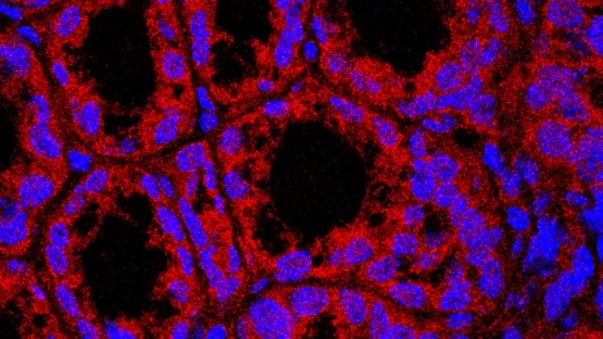
The DELTA method operates by tagging a synaptic protein of interest in the mouse brain using a vibrant Janelia Fluor dye. To study how synaptic connections evolve during learning, researchers trained mice to associate distinct visual cues with a reward of water. Post-labeling, the mice were divided into two groups; one experienced random water rewards at both cues, while the other learned that only one cue would lead to a reward. After observing significant behavioral differences, the scientists performed a second labeling with a different dye, allowing them to track changes in the same synaptic protein, known as GluA2.
Key Findings
Through imaging the entire brain over several days, researchers identified regions where synaptic proteins shifted in response to the modified learning task. Excitingly, they discovered that such learning was linked to specific changes in GluA2 levels in key brain areas. Moreover, they also compared the effects of normal environments versus enriched environments filled with toys and social interaction—finding that enrichment led to widespread GluA2 changes throughout the brain.
Future Research Directions
By revealing these extensive brain-wide modifications, DELTA sets the stage for future investigations into the cellular and molecular frameworks supporting learning and memory. The research team is already aiming to enhance DELTA further, hoping to determine the precise timing of protein changes during the learning process.
Collaboration and Sharing Knowledge
Nelson Spruston, one of the project's leading scientists, lauds the collaborative spirit at Janelia, noting that the success of DELTA stemmed from pooling expertise across disciplines, including chemistry, imaging, and genetics. Partnerships extended beyond Janelia, incorporating insights from experts at Northwestern University to validate the technique.
A New Era of Neuroscience
Now, the team is sharing DELTA with researchers worldwide through Janelia’s Visiting Scientist Program, enabling them to track synaptic changes in their own studies.
Conclusion
This project epitomizes the essence of Janelia,” Spruston states. “We achieved something entirely unprecedented by leveraging collective expertise, pushing the boundaries of what we know about the brain.” As scientists continue to unravel the complexities of brain function through DELTA, we stand on the brink of transformative discoveries in our understanding of how learning and memory operate at the molecular level. Buckle up for the ride into the brain's intricate workings!


 Brasil (PT)
 Canada (EN)
 Chile (ES)
 Česko (CS)
 대한민국 (KO)
 España (ES)
 France (FR)
 Hong Kong (EN)
 Italia (IT)
 日本 (JA)
 Magyarország (HU)
 Norge (NO)
 Polska (PL)
 Schweiz (DE)
 Singapore (EN)
 Sverige (SV)
 Suomi (FI)
 Türkiye (TR)
 الإمارات العربية المتحدة (AR)