Revolutionary Handheld Scanner Redefines Cancer and Arthritis Diagnosis
2024-09-30
Introduction
In a groundbreaking innovation, researchers at University College London (UCL) have unveiled a handheld scanner that can produce stunningly detailed 3D images using photoacoustic tomography (PAT)—a technique that could revolutionize the early diagnosis of cancer, cardiovascular diseases, and arthritis. This remarkable advancement, detailed in a recent publication in Nature Biomedical Engineering, holds the promise of significantly enhancing clinical imaging practices.
Imaging Process Revolutionized
Traditional photoacoustic imaging methods could take over five minutes to generate a usable image, requiring patients to remain completely still—a challenging feat in many cases. However, UCL's state-of-the-art scanner dramatically accelerates this process by simultaneously scanning multiple points of tissue, cutting down imaging time to mere seconds and capturing incredibly fine details of blood vessels, even in motion-prone situations.
Expert Insights
Paul Beard, PhD, a professor of medical physics and biomedical engineering at UCL, emphasizes the significance of this breakthrough. “This technology is 100 to 1,000 times faster than previous solutions," Beard explains. "It enables clinicians to visualize dynamic physiological events without motion-induced blurring, revealing unprecedented image quality.”
Implications for Arthritis Patients
This leap in technology carries vital implications for patients with conditions like inflammatory arthritis, requiring scans of numerous joint areas like the fingers. The older generation of PAT scanners might take nearly an hour—too daunting for elderly or frail patients—while the new device can complete the process in just a few minutes.
Clinical Trials
The clinical trials for this new scanner have commenced, showcasing its potential through experiments on 10 patients with varying medical conditions, including type-2 diabetes and breast cancer, as well as healthy individuals. In a striking demonstration, physicians were able to visualize microvascular deformities in diabetic patients' feet and detect inflammation linked to the malignancy in breast cancer cases.
Findings on Diabetes and Microvascular Health
Andrew Plumb, PhD, an associate professor of medical imaging at UCL, highlighted the importance of these findings: “For diabetes patients, low blood flow in extremities can occur due to tiny blood vessel damage—previously invisible until now.” The UCL team's scanner distinguished between healthy and damaged vessels in study participants, paving the way for earlier intervention and prevention of severe complications.
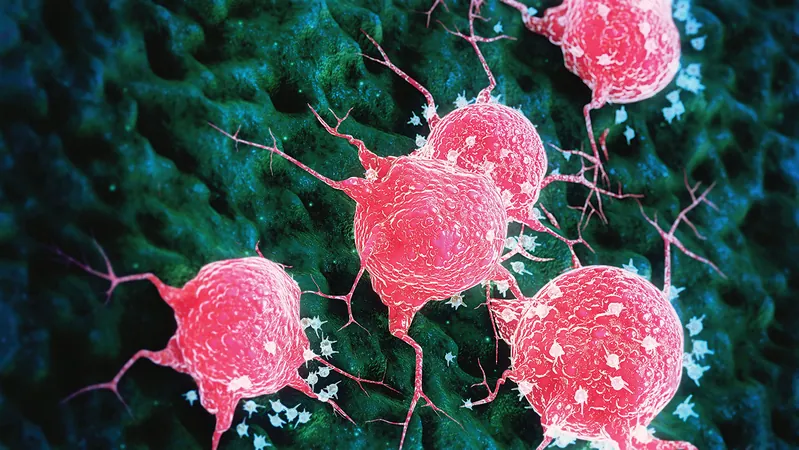
Cancer Care Applications
Moreover, the potential applications in cancer care are enormous. Scanner co-developer Nam Huynh, PhD, noted, “Photoacoustic imaging offers an accessible method for tumor detection and monitoring. It enhances surgical precision by providing surgeons with greater insight into tumor boundaries, minimizing the chances of residual cancerous tissue and thereby reducing recurrence risk.”
Future Research and Implications
As the UCL team plans additional studies with larger patient groups, this pioneering scanner emerges as a pivotal stride towards reshaping how we diagnose and understand diseases. While success in medical imaging is an extraordinary milestone, the ongoing research aims to uncover its practicality in various clinical settings.
Conclusion
This cutting-edge technology could mean the difference between a timely diagnosis and late-stage disease intervention, marking a hopeful turn in medical innovation. Stay tuned as this incredible scanner might change the face of healthcare as we know it!
 Brasil (PT)
 Canada (EN)
 Chile (ES)
 España (ES)
 France (FR)
 Hong Kong (EN)
 Italia (IT)
 日本 (JA)
 Magyarország (HU)
 Norge (NO)
 Polska (PL)
 Schweiz (DE)
 Singapore (EN)
 Sverige (SV)
 Suomi (FI)
 Türkiye (TR)