Revolutionary Crystal Could Redefine Cancer and Heart Disease Scans
2025-09-15
Author: Arjun
Imagine a medical scan that delivers crystal-clear images of your heart or brain in mere minutes, while significantly reducing radiation exposure and costs for hospitals. This futuristic vision is on the brink of becoming a reality thanks to groundbreaking new detector technology.
Researchers from Northwestern University (NWU) and Soochow University have created a pioneering perovskite-based gamma-ray detector for nuclear medicine. This innovative device captures signals with unprecedented clarity, potentially revolutionizing how doctors diagnose heart disease, cancer, and other hidden disorders.
The Limitations of Existing Detectors
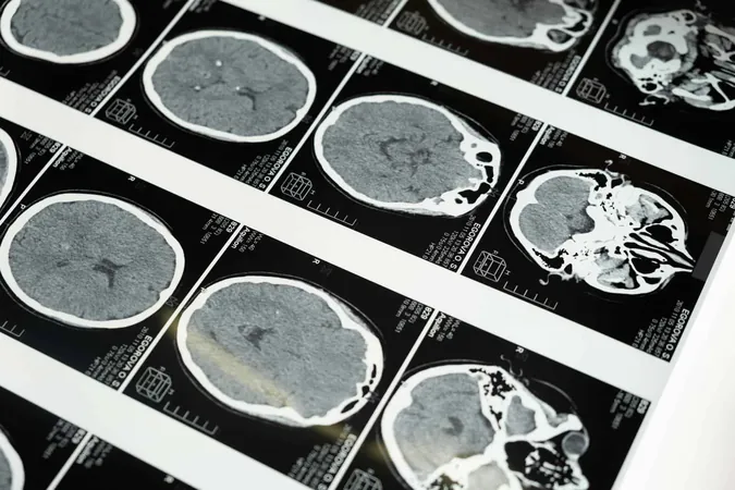
In conventional nuclear medicine scans, patients receive a tiny amount of a safe, short-lived radioactive tracer. This tracer targets specific body parts, emitting gamma rays detected by external cameras that create 3D images. Tools like SPECT (single-photon emission computed tomography) have been essential for monitoring blood flow, heart function, and tumor detection. However, existing detectors fall short in terms of performance and cost.
While advanced detectors made from cadmium zinc telluride (CZT) offer high precision, they also come with hefty price tags, sometimes exceeding millions of dollars. Conversely, sodium iodide (NaI) detectors, though more affordable, produce unclear, blurry images, inhibiting widespread access to top-tier nuclear imaging.
The Game-Changing Potential of Perovskite Detectors
Back in 2013, researchers first recognized that perovskites, known primarily for their role in solar energy, also hold immense potential in radiation detection. In this latest study, they successfully crafted large, high-quality cesium lead bromide (CsPbBr₃) crystals into pixelated sensors, designed like smartphone camera pixels for precise radiation capture.
This system achieved remarkable energy resolution, hitting 2.5% at 141 kiloelectronvolts, the energy typical of the most widely used clinical radiotracer, technetium-99m. Even at higher energy levels, such as 662 kiloelectronvolts, the system achieved a 1.0% resolution—the best reported for perovskite detectors yet.
In practical imaging tests, the device showcased its ability to discern radioactive sources just a few millimeters apart, accurately capturing fine anatomical details and even detecting faint signals.
A Practical Innovation for Health Care
The implications of this research are significant for healthcare. If fully developed, perovskite detectors could make nuclear scans significantly sharper, quicker, more affordable, and less radiation-intensive. This means patients would spend less time in scanners and receive clearer, more reliable results.
Hospitals with limited budgets could finally access cutting-edge imaging technology, bridging the divide between elite research institutions and community clinics. As Kanatzidis, the lead study author, stated, "High-quality nuclear medicine shouldn’t be limited to hospitals that can afford expensive equipment. With perovskites, we can deliver faster, safer scans to many more patients worldwide. The end goal is improved scans, diagnoses, and patient care."
This achievement stands as a milestone for both physics and materials science, marking the first successful demonstration of single-photon gamma-ray imaging using perovskites, showcasing their transition from laboratory novelty to practical application. A new NWU spinout, Actinia Inc., is already collaborating with medical device companies to introduce this transformative technology into healthcare.


 Brasil (PT)
 Canada (EN)
 Chile (ES)
 Česko (CS)
 대한민국 (KO)
 España (ES)
 France (FR)
 Hong Kong (EN)
 Italia (IT)
 日本 (JA)
 Magyarország (HU)
 Norge (NO)
 Polska (PL)
 Schweiz (DE)
 Singapore (EN)
 Sverige (SV)
 Suomi (FI)
 Türkiye (TR)
 الإمارات العربية المتحدة (AR)