Revolutionary Cell Imaging: How AI is Transforming Traditional Techniques into a Game-Changer for Live-Cell Analysis!
2025-01-17
Author: Mei
Revolutionary Cell Imaging: How AI is Transforming Traditional Techniques into a Game-Changer for Live-Cell Analysis!
An innovative breakthrough from researchers at POSTECH has the potential to revolutionize the world of cell imaging. Published in Nature Communications, their technology deftly overcomes the limitations of conventional imaging methods, ensuring stable and highly accurate cellular visualization.
Conventional methods vs Mid-Infrared Photoacoustic Microscopy
For decades, confocal fluorescence microscopy (CFM) has been a cornerstone in the life sciences for its ability to produce stunning high-resolution images of cells. However, the technique's reliance on fluorescent staining raises serious concerns about photobleaching and phototoxicity, which can harm the very cells researchers aim to study. In contrast, mid-infrared photoacoustic microscopy (MIR-PAM) offers a label-free imaging alternative that safeguards cell integrity. The catch? Its longer wavelengths inadvertently restrict spatial resolution, making it tricky to visualize intricate cellular structures.
Groundbreaking Imaging Method: Explainable Deep Learning (XDL)
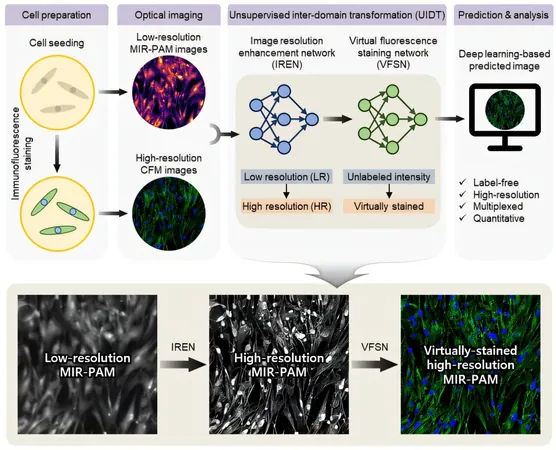
To tackle these challenges head-on, the POSTECH research team introduced a groundbreaking imaging method leveraging explainable deep learning (XDL). This cutting-edge approach transforms low-resolution, label-free MIR-PAM images into high-resolution, virtually stained visuals that imitate the quality of images from CFM. A standout feature of XDL is its remarkable transparency. Unlike traditional AI models, XDL allows researchers to see the transformation process, ensuring reliable and accurate results.
Two-Phase Imaging Process
The researchers implemented a single-wavelength MIR-PAM system and devised a sophisticated two-phase imaging process. The first phase, known as the Resolution Enhancement phase, converts low-resolution MIR-PAM images into crisp, high-resolution counterparts, revealing complex cellular structures such as nuclei and filamentous actin. The second phase, dubbed Virtual Staining, creates virtually stained images devoid of fluorescent dyes—effectively eliminating the risks that come with traditional staining while delivering imagery of CFM caliber.
Significance of the Research
This pioneering technology not only ensures high-resolution, virtually stained cellular imaging but does so without compromising cell health, presenting a robust new instrument for live-cell analysis and advanced biological research.
Expert Insights
Professor Chulhong Kim emphasized the significance of their work, stating, "We have developed a cross-domain image transformation technology that bridges the physical limitations of different imaging modalities, offering complementary benefits. The XDL approach has significantly enhanced the stability and reliability of unsupervised learning."
Adding to this sentiment, Professor Jinah Jang remarked, "This research unlocks new possibilities for multiplexed, high-resolution cellular imaging without labeling. It holds immense potential for applications in live-cell analysis and disease model studies.”
Conclusion and Future Prospects
With this revolutionary advancement, researchers are poised to explore the inner workings of cells like never before, paving the way for groundbreaking discoveries in areas such as cancer research, cellular biology, and regenerative medicine. Stay tuned as the future of biological research gets ready to embark on this exciting new journey!


 Brasil (PT)
 Canada (EN)
 Chile (ES)
 Česko (CS)
 대한민국 (KO)
 España (ES)
 France (FR)
 Hong Kong (EN)
 Italia (IT)
 日本 (JA)
 Magyarország (HU)
 Norge (NO)
 Polska (PL)
 Schweiz (DE)
 Singapore (EN)
 Sverige (SV)
 Suomi (FI)
 Türkiye (TR)
 الإمارات العربية المتحدة (AR)