Revolutionary AI Breakthrough: Clearer Images of Biological Samples Without Extra Equipment!
2025-01-29
Author: Wei
Introduction
Biologists have long faced a major hurdle in microscopy: as they delve deeper into biological samples, the clarity of their images suffers due to light distortion. Even the tiniest samples, such as worm embryos or tissue sections measuring mere tens of microns, can become obscured and fuzzy when viewed with traditional microscopes.
The Challenge of Light Distortion
To combat this issue, many researchers have relied on adaptive optics technology, a sophisticated solution designed to correct these distortions. However, the required investment of time, money, and specialized knowledge has restricted this technology's accessibility, leaving many biology labs unable to utilize it.
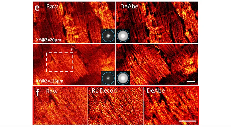
A Breakthrough from HHMI's Janelia Research Campus
Enter the innovative team from HHMI's Janelia Research Campus. Led by members of the esteemed Shroff Lab, they’ve pioneered a groundbreaking AI-driven technique that captures ultra-clear images of thick biological samples—without the need for adaptive optics or additional hardware. This remarkable achievement has the potential to democratize high-quality microscopy.
How It Works
The Shroff Lab team achieved this by first developing a model to understand and replicate the degradation effect that occurs when a microscope images deeper layers of a sample. They then used untainted near-side images and artificially distorted them, akin to the deeper images. With this experimental setup, they trained a neural network to reverse the distortions, resulting in enhanced clarity throughout the entire depth of the sample.
Enhanced Accuracy in Biological Assessments
Not only does this deep learning-based method yield stunningly clear images, but it has also significantly improved the accuracy of biological assessments. Researchers can now more effectively count cells in worm embryos, trace intricate blood vessels and pathways in whole mouse embryos, and examine critical cellular structures like mitochondria in liver and heart samples.
Simplicity and Accessibility
What makes this technique especially revolutionary is its simplicity: all it requires is a standard microscope, a computer equipped with a graphics card, and a brief tutorial to get started. This contrasts sharply with traditional adaptive optics, making advanced imaging techniques attainable for a broader range of biological research labs.
Ongoing Research and Future Potential
The Shroff Lab is already applying this transformative method to their ongoing research on worm embryos and is in the process of refining their model to accommodate less uniform samples. This marks a significant leap forward in biological imaging—one that could reshape how scientists visualize and understand complex biological systems.
Conclusion
As biological research continues to advance, this AI-enhanced imaging method could unlock new possibilities in the study of health, disease, and regenerative biology. Are we on the brink of a microscopy revolution? Stay tuned as this innovative technology unfolds!


 Brasil (PT)
 Canada (EN)
 Chile (ES)
 Česko (CS)
 대한민국 (KO)
 España (ES)
 France (FR)
 Hong Kong (EN)
 Italia (IT)
 日本 (JA)
 Magyarország (HU)
 Norge (NO)
 Polska (PL)
 Schweiz (DE)
 Singapore (EN)
 Sverige (SV)
 Suomi (FI)
 Türkiye (TR)