Revolutionary 3D Imaging Technique Unveils Enzyme Activity Secrets in Whole Organs!
2025-04-28
Author: Arjun
Uncovering the Hidden World of Enzyme Activity
Imagine peering deep into an organ and seeing how enzymes perform their vital roles in real time. Thanks to groundbreaking advances in 3D imaging, researchers can now visualize enzyme activity with astonishing clarity in whole organs! This innovation, led by a team from the University of Tokyo, harnesses specially designed probe molecules that light up when activated by specific enzymes.
The Power of High-Resolution Imaging
Published in the prestigious journal Angewandte Chemie International Edition, this study showcased how the technique allowed visualization of aminopeptidase N (APN) activity and the impact of drug inhibitors in mouse kidneys. Enzymes like APN are essential for various bodily functions, and abnormalities in their activity can signal serious health issues. Yet, until now, capturing the intricate details of enzyme activity across different regions of an organ has been a significant challenge.
Breaking Through the Limitations of Traditional Imaging
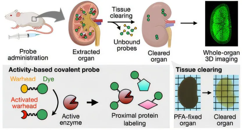
Traditional imaging methods often fall short because fluorescent light struggles to penetrate thick tissue samples. The clever solution? A process called "clearing" that renders organs transparent while keeping their structure intact. However, this process usually washes away smaller fluorescent probes, making it difficult to retain the crucial information they provide.
A Game-Changing Method for Enzyme Imaging
The innovative approach developed by Shinsuke Sando and his team changes the game entirely. By utilizing a unique probe that contains a fluorescent dye, an anchoring unit, and an amino acid group, they can effectively map enzyme activity. In the presence of active APN, the probe is modified in such a way that it clings tightly to nearby proteins, ensuring the fluorescent signal remains intact during the clearing process.
Revealing APN Activity in 3D
Their groundbreaking method enabled the researchers to produce stunning 3D images of APN activity in the mouse kidneys, revealing not just overall activity but also nuanced differences in enzyme behavior within individual tubular structures. This unprecedented level of detail offers new insights into how enzymes function at a micro-level.
Implications for Drug Development
The team didn’t stop there; they also explored how different APN inhibitors affected enzyme activity. By comparing the experimental antitumor drug actinonin with another inhibitor, they discovered significant differences in how these drugs suppressed fluorescence, hinting at varying absorption and metabolism rates. This knowledge could be crucial for future drug development and tailored therapies.
The Future of Enzyme Research Looks Bright
With this revolutionary technique, the potential for deeper understanding of enzyme functions and pathological conditions is more promising than ever. As researchers continue to uncover the complexities of enzyme activities within whole organs, we move closer to breakthroughs that could transform the field of medicine!

 Brasil (PT)
 Canada (EN)
 Chile (ES)
 Česko (CS)
 대한민국 (KO)
 España (ES)
 France (FR)
 Hong Kong (EN)
 Italia (IT)
 日本 (JA)
 Magyarország (HU)
 Norge (NO)
 Polska (PL)
 Schweiz (DE)
 Singapore (EN)
 Sverige (SV)
 Suomi (FI)
 Türkiye (TR)
 الإمارات العربية المتحدة (AR)