Revolutionary 12-Minute MRI Technique Promises Early Detection of Brain Diseases
2025-07-02
Author: Arjun
Unlocking Brain Secrets at Lightning Speed
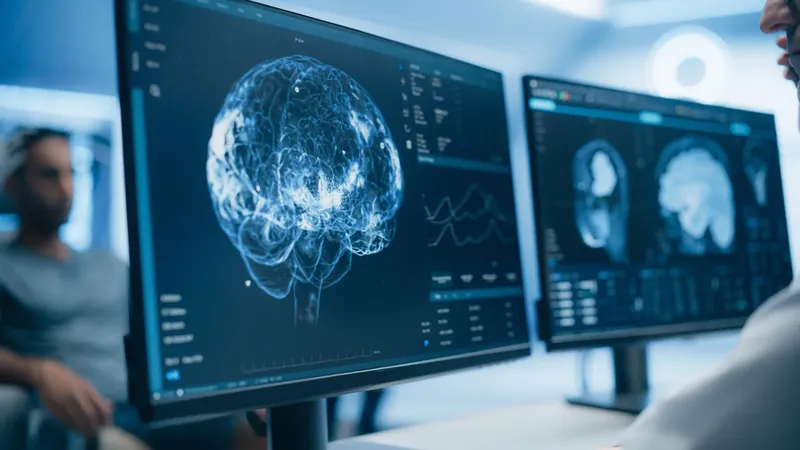
A groundbreaking advancement in medical imaging promises to change how we diagnose brain diseases by mapping brain chemistry in just 12 minutes. Spearheaded by Zhi-Pei Liang, a leading professor in electrical and computer engineering at the University of Illinois, this innovative technology enhances traditional MRI methods to provide critical insights into brain metabolism.
Metabolism: The Key to Understanding Brain Health
For over four decades, MRI technology has been instrumental in deciphering the complex workings of the brain. While conventional MRI excels in visualizing brain structures and fMRI highlights blood flow changes linked to neural activity, neither could assess the brain's metabolic processes—crucial for understanding both function and disease progression.
Postdoctoral researcher Yibo Zhao, the study's lead author, explains, "Metabolic shifts often precede visible structural or functional changes in the brain, making metabolic imaging a game changer for early diagnosis and potential interventions." The new approach focuses on measuring signals from brain metabolites and neurotransmitters, enhancing the diagnostic capabilities of MRI.
Conquering Challenges in Brain Imaging
Previous attempts at Magnetic Resonance Spectroscopic Imaging (MRSI) faced hurdles like lengthy scan times and interference from noise, complicating the interpretation of neurotransmitter signals. However, Liang’s team has cracked this dilemma by combining ultrafast data acquisition techniques with advanced machine learning algorithms, achieving remarkable speed and resolution.
Real-World Applications: Diagnosing Conditions Faster
In clinical tests, the researchers successfully demonstrated the technique's effectiveness on various patient groups. In healthy individuals, they mapped diverse metabolic patterns across brain regions, revealing that activity levels aren’t uniform across the brain. For patients with brain tumors, notable metabolic changes were detected—such as elevated levels of choline and lactate—even when tumors appeared indistinguishable in standard MRI scans.
Additionally, subjects with multiple sclerosis showed molecular changes related to inflammation and reduced neuronal activity up to 70 days before they could be identified using conventional imaging.
Transforming the Future of Neurological Treatment
The implications of this high-speed, high-resolution imaging technique are vast. As Liang shares, this technology not only allows clinicians to track metabolic changes over time but also supports personalized medicine by tailoring treatments to individual metabolic profiles. With the healthcare landscape shifting towards precision medicine, this breakthrough could meet an urgent need for noninvasive metabolic imaging in clinical environments.
A Nod to the Past, a Vision for the Future
Reflecting on the journey, Liang notes the influence of the late Professor Paul Lauterbur, a Nobel Prize winner who pioneered MRI technology. "While he envisioned the potential for fast metabolic imaging, realizing it in a clinical setting has been immensely challenging. Our advancement aims to fulfill that promise," Liang concludes.
A Bright Future Ahead
This transformative research, backed by the Arnold and Mabel Beckman Foundation, signals a new era in brain health diagnostics, allowing for quicker, more accurate interventions that could save lives through earlier detection of neurological diseases.

 Brasil (PT)
 Canada (EN)
 Chile (ES)
 Česko (CS)
 대한민국 (KO)
 España (ES)
 France (FR)
 Hong Kong (EN)
 Italia (IT)
 日本 (JA)
 Magyarország (HU)
 Norge (NO)
 Polska (PL)
 Schweiz (DE)
 Singapore (EN)
 Sverige (SV)
 Suomi (FI)
 Türkiye (TR)
 الإمارات العربية المتحدة (AR)