Groundbreaking Whole-Brain Atlas Unlocks Secrets of Motor Control
2024-12-24
Author: Nur
In a major breakthrough for neuroscience, researchers at St. Jude Children’s Research Hospital have unveiled an innovative whole-brain atlas that sheds light on how the brain orchestrates muscle movement. While it’s well-known that the brain sends signals to motor neurons to facilitate muscle contractions, the intricate pathway these signals take—specifically through a diverse group of spinal interneurons—has largely remained a mystery.
Dr. Jay Bikoff, the study's lead author and expert in developmental neurobiology, emphasized the complexity of this connection. “For decades, our understanding of the motor system has highlighted its distributed nature, but the critical link through the spinal cord and its diverse interneurons was underexplored,” he explained. These interneurons serve as a crucial intermediary, modulating the activity of motor neurons and, consequently, influencing muscle movements.
The Hidden Terrain of Interneurons: A Vast Network to Understand
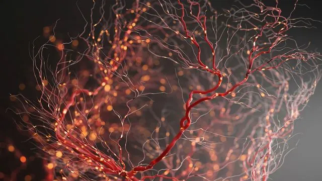
Until now, the relationship between brain regions responsible for motor control and specific neurons in the spinal cord has been akin to searching in the dark. Interneurons come in numerous varieties, complicating efforts to pinpoint how they interact with motor neurons. “Exploring this network is like untangling a ball of lights—and we’re dealing with over 3 billion years of evolutionary complexity,” remarked co-first author Dr. Anand Kulkarni.
To bridge this knowledge gap, the research team utilized a genetically modified strain of the rabies virus, lacking a vital surface protein that usually enables it to jump between neurons. This clever tactic allowed the virus to remain at its point of origin while identifying connections to specific interneurons. Using a fluorescent tracking system, researchers could trace the origin of inputs to the V1 interneurons, which are critical for motor output but previously shrouded in ambiguity.
Mapping the Neural Landscape: A New Frontier
The groundbreaking work resulted in a detailed three-dimensional interactive atlas of the brain’s connections to spinal interneurons. Researchers employed serial two-photon tomography to create this visual representation, effectively transforming the brain into hundreds of micron-thick slices revealing the interconnectedness of neurons.
“This atlas not only maps out the V1 interneurons but also allows us to generate hypotheses about the brain regions influencing motor function. It’s a hypothesis-generating engine for future research,” Bikoff explained. With the data freely accessible through a web-based platform, both scientists and students across the globe can now delve into this exhilarating domain of neuroscience.
Implications for Future Research and Therapies
This groundbreaking atlas could pave the way for more in-depth investigations into various disorders related to motor control, such as Parkinson's disease and spinal cord injuries. Understanding the precise pathways and networks involved in motor function significantly enhances our capability to develop targeted therapies that could help restore movement in affected individuals.
As Bikoff concluded, “This research provides a crucial framework not only for understanding the neural basis of movement but also for the development of therapeutic strategies aimed at treating movement disorders. The implications of our findings could potentially transform the way we approach neurorehabilitation.”
This remarkable advancement marks a significant step forward in neuroscience, offering new insights into how our brains control movement and opening up exciting avenues for future exploration and innovation.


 Brasil (PT)
 Canada (EN)
 Chile (ES)
 Česko (CS)
 대한민국 (KO)
 España (ES)
 France (FR)
 Hong Kong (EN)
 Italia (IT)
 日本 (JA)
 Magyarország (HU)
 Norge (NO)
 Polska (PL)
 Schweiz (DE)
 Singapore (EN)
 Sverige (SV)
 Suomi (FI)
 Türkiye (TR)
 الإمارات العربية المتحدة (AR)