Groundbreaking Study Uncovers How Your Brain Controls Blood Flow
2025-07-21
Author: Siti
Revolutionary Discovery in Brain Function
A team of researchers at the Center for Neuroscience Imaging Research, part of the Institute for Basic Science (IBS), has announced a groundbreaking discovery regarding how the brain regulates its blood flow. This insight could reshape our understanding of functional magnetic resonance imaging (fMRI) and its interpretations.
The Role of Inhibitory Neurons Revealed
At the heart of this study are somatostatin-expressing interneurons (SST neurons)—a specific type of inhibitory neuron. While about 15% of all neurons in the brain are inhibitory, their role in controlling blood volume has mostly gone unexplored, overshadowed by the focus on excitatory neurons.
How Neurons and Astrocytes Work Together
Published in *Nature Communications*, the research reveals how inhibitory neurons collaborate with astrocytes, supportive brain cells, to regulate the dilation of blood vessels precisely. The scientists developed innovative mouse models (like SST-ChR2, SST-hM4Di) to study this interaction more closely, employing cutting-edge techniques such as optogenetics and chemogenetics.
To track these interactions, they used calcium imaging to visualize astrocyte responses, electrophysiology for neural signal measurement, and advanced imaging methods like ultra-high-field fMRI to observe blood volume changes with remarkable precision.
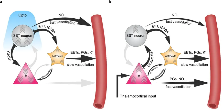
Unveiling the Two-Step Mechanism
Through their experiments, the team discovered a two-step vasodilation mechanism. Initially, activated SST neurons release nitric oxide (NO), a potent vasodilator, prompting a swift expansion of nearby blood vessels. This is succeeded by a slower phase where astrocytes trigger localized dilation, particularly during extended sensory stimulation.
Significance for Brain Imaging
Crucially, silencing SST neurons resulted in a loss of layer-specific precision in fMRI blood volume signals, directly associating neural activity with spatial specificity in imaging data. "This two-step interaction between inhibitory neurons and astrocytes illuminates why high-resolution fMRI can discern activities across different brain cortex layers," the researchers noted.
Implications for Neurological Health
This research not only enhances our understanding of neurovascular coupling but also opens new pathways for exploring neurological and psychiatric disorders where SST neuron function is compromised, including Alzheimer's disease, depression, and autism. The team plans to investigate these conditions further using disease-model mice to see how disruptions within this neuron-astrocyte-vascular system impact blood volume, brain function, and fMRI signals.
A New Era in Brain Imaging and Treatments
Ultimately, this pioneering research lays a strong foundation for refining brain imaging accuracy, potentially leading to improved diagnostic and therapeutic strategies for various brain disorders.


 Brasil (PT)
 Canada (EN)
 Chile (ES)
 Česko (CS)
 대한민국 (KO)
 España (ES)
 France (FR)
 Hong Kong (EN)
 Italia (IT)
 日本 (JA)
 Magyarország (HU)
 Norge (NO)
 Polska (PL)
 Schweiz (DE)
 Singapore (EN)
 Sverige (SV)
 Suomi (FI)
 Türkiye (TR)
 الإمارات العربية المتحدة (AR)