Groundbreaking Microscopy Captures Live Cell Wall Construction in Plants, Paving the Way for Agricultural Innovations
2025-03-25
Author: Ming
In an exciting breakthrough, researchers at Rutgers University-New Brunswick have achieved a historic feat—capturing live, continuous images of cellulose synthesis in living plant cell walls for the very first time. This groundbreaking study, which spanned over 24 hours, has profound implications for enhancing food production and developing sustainable biofuels.
This unprecedented ability to visualize the dynamic process of cell wall formation expands our understanding of how plants build their structural framework. "We are witnessing the direct real-time visualization of cellulose being synthesized and self-assembling into a complex network," explained Associate Professor Sang-Hyuk Lee from the Department of Physics and Astronomy. He emphasizes how this study reveals new insights into the fundamental processes guiding cellular organization through simple physical mechanisms like diffusion.
The research brings together three diverse laboratories at Rutgers, marking six years of collaboration among experts in biophysics, bioengineering, and plant biology from the School of Arts and Sciences, the School of Engineering, and the School of Environmental and Biological Sciences. Together, they harnessed cutting-edge technology to pioneer an advanced imaging technique known as total internal reflection fluorescence microscopy.
Cellulose, a carbohydrate and the most abundant biopolymer on Earth, serves as the primary structural component of plant cell walls. Its importance extends far beyond botany, with numerous industrial applications that range from paper and textiles to food thickening agents and filtration processes.
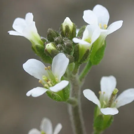
To capture their groundbreaking findings, the team used protoplasts, which are plant cells devoid of their walls, from the domestically significant Arabidopsis plant—a staple organism used extensively in plant research. The results revealed an astonishing spectacle: seemingly chaotic sprouting cellulose fiber filaments gradually assembled into a well-organized network, defying the traditional perceptions held by many scientists that cellulose is synthesized in a more orderly fashion.
"I was astounded by the emergence of these structured networks from what I initially expected to be a more orderly process," said Lee, who also holds a position at the Institute for Quantitative Biomedicine.
This discovery not only satisfies scientific curiosity but opens a gateway to crucial applications in genetics and biotechnology. According to Distinguished Professor Eric Lam, one of the authors, "This discovery equips us to explore the genes involved in cellulose biosynthesis, allowing us to create better plants that can effectively capture carbon and enhance stress tolerance against drought and disease."
Moreover, Associate Professor Shishir Chundawat highlighted the relevance of this research in the context of sustainable engineering, aiming to produce valuable bioproducts that can benefit society. "My lifelong fascination with how plants harness sunlight into forms like cellulose inspires our drive to understand biomass production further."
This innovative approach also allowed researchers to tag emerging cellulose structures with a special fluorescent protein dye, enabling detailed observation without the risk of damaging the delicate cells. Lee led the imaging efforts, while Chundawat's team made significant contributions through their work on protein engineering, ensuring that the cellulose filaments were visible under the microscope.
With the technology now in place, Rutgers scientists foresee an era of advanced plant research that could significantly influence sustainable agricultural practices and bioproduct development, ultimately enhancing global food security and reducing reliance on fossil fuels. The future of plant biology is bright, and this breakthrough may just be the spark that ignites a revolution in how we understand and utilize plant life.


 Brasil (PT)
 Canada (EN)
 Chile (ES)
 Česko (CS)
 대한민국 (KO)
 España (ES)
 France (FR)
 Hong Kong (EN)
 Italia (IT)
 日本 (JA)
 Magyarország (HU)
 Norge (NO)
 Polska (PL)
 Schweiz (DE)
 Singapore (EN)
 Sverige (SV)
 Suomi (FI)
 Türkiye (TR)
 الإمارات العربية المتحدة (AR)