Groundbreaking Discovery Unveils How Cells Process Glucose: A New Metabolic Map!
2025-07-21
Author: Wei
A Scientific Breakthrough in Metabolic Mapping
In an unprecedented achievement, researchers from Vanderbilt University and the University of California, San Diego, have crafted a detailed metabolic "map" showing how cells process glucose, unveiling a hidden realm where organelles work together in response to nutrient influx.
Published in Nature Communications, this revolutionary study changes the landscape of glucose metabolism visualization at the single-cell level, providing groundbreaking methods and insights that can help examine disruptions in metabolic processes related to diseases such as diabetes, obesity, cancer, aging, and neurodegeneration.
Integrating Cutting-Edge Techniques for Remarkable Insights
Rafael Arrojo e Drigo, an assistant professor of molecular physiology and biophysics and the lead author of the study, stated, "We are pioneering the integration of various microscopy techniques to trace the journey of glucose atoms across different biological scales—from entire animals down to individual organelles and their intricate architectures."
This innovation opens the door for a new research program aimed at understanding how nutrient metabolism is organized within the structured domains of cells and tissues, which is crucial for the precise regulation of organ function within overall bodily physiology.
A Shift from Bulk Analyses to Single-Cell Insights
Traditionally, knowledge about cellular nutrient processing comes from bulk metabolomics, which analyzes a collective set of molecules in biological samples without accounting for their specific spatial context within cells. Arrojo e Drigo highlighted the limitation of these bulk methods, stating they fail to capture the spatial dynamics of metabolism on a single-cell level.
This insight prompted a cross-disciplinary team from Vanderbilt, Vanderbilt University Medical Center, and UCSD to fuse stable isotope tracing, multi-scale microscopy, and AI image analysis to create a comprehensive glucose metabolites map spanning across animal, tissue, cellular, and organelle levels.
Revealing the Complex Interactions of Organelles
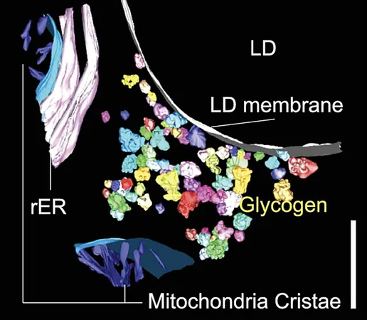
Their findings revealed the spatial organization of glucose metabolites, tracing their incorporation into glycogen, lipid droplets, and other cellular components in live mice. A key revelation was a newly identified structural interaction between lipid droplets and glycogen synthesis, showcasing the dynamic relationship between these two critical elements.
The researchers also documented how the interaction points between mitochondria and the endoplasmic reticulum—organelle powerhouses crucial for energy production—adapt fluidly based on fluctuating blood glucose levels, forming a part of a wider organelle network that orchestrates metabolic responses within cells.
Looking Ahead: The Future of Metabolic Research
This breakthrough owes much to Vanderbilt's collaborative research environment and the multi-modal imaging capabilities of the National Center for Microscopy and Imaging Research (NCMIR). As Arrojo e Drigo noted, the team's goal is to further explore how nutrient spatial organization within cells affects metabolic health and the development of diseases.
The research team was co-led by Christopher Acree and Aliyah Habashy from the Arrojo e Drigo lab, with contributions from scientists across various departments at Vanderbilt and UCSD, enhancing the interdisciplinary collaboration that made this breakthrough possible.


 Brasil (PT)
 Canada (EN)
 Chile (ES)
 Česko (CS)
 대한민국 (KO)
 España (ES)
 France (FR)
 Hong Kong (EN)
 Italia (IT)
 日本 (JA)
 Magyarország (HU)
 Norge (NO)
 Polska (PL)
 Schweiz (DE)
 Singapore (EN)
 Sverige (SV)
 Suomi (FI)
 Türkiye (TR)
 الإمارات العربية المتحدة (AR)