Gold Rush in Cardiology: Revolutionary Imaging Breakthrough Using Gold Nanoparticles!
2024-10-28
Author: Rajesh
Gold Rush in Cardiology: Revolutionary Imaging Breakthrough Using Gold Nanoparticles!
In an exciting development for heart disease diagnostics, researchers from the University of Ottawa have unveiled a groundbreaking contrast agent that could redefine cardiac imaging capabilities. This cutting-edge advancement utilizes gold nanoparticles in a medical imaging technique known as intravascular optical coherence tomography (IV-OCT), significantly enhancing the ability to diagnose heart conditions more accurately.
Led by Associate Professor Adam J. Shuhendler, the research team successfully created gold superclusters (AuSC) tailored specifically for the near-infrared light employed in IV-OCT. These innovative superclusters consist of densely packed gold nanoparticles, which dramatically improve light scattering and, consequently, imaging clarity. Their research, titled “NIR-II Scattering Gold Superclusters for Intravascular Optical Coherence Tomography Molecular Imaging,” was published in the prestigious journal Nature Nanotechnology.
Shuhendler explains, "We've discovered a simple and efficient method to produce these gold superclusters, and we can fine-tune them for optimal performance in IV-OCT imaging." By coating the gold superclusters with a specialized polymer, the team ensured their stability while enabling the attachment of targeting molecules, thus enhancing specificity in imaging.
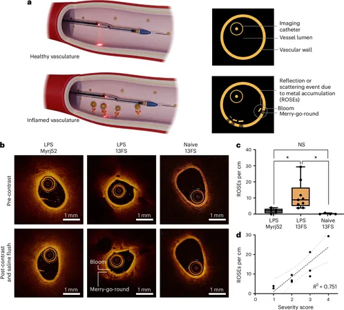
Central to their study was P-selectin, a known marker of inflammation in blood vessels. The newly developed contrast agent, dubbed AuSC@(13FS)2, demonstrated robust binding capabilities to P-selectin in laboratory tests and significantly improved IV-OCT imaging results in rat subjects with inflamed blood vessels. This marks a pivotal advancement in the quest for detailed and specific imaging of cardiovascular issues.
One of the standout advantages of this innovative agent is its compatibility with existing IV-OCT procedures in clinical settings, allowing for seamless integration without significant modifications to current practices. Remarkably, when AuSC@(13FS)2 interacted with inflamed blood vessels, it generated distinct reflections within the IV-OCT images — reflections akin to those observed when using stents, paving the way for clearer diagnostics.
"Our new contrast agent has the potential to pave the way for more personalized treatment approaches in heart disease," Shuhendler asserts. This cutting-edge technology may empower physicians to detect heart diseases at earlier stages and more accurately assess associated risks by providing nuanced insights into blood vessel health.
The research also uncovered a direct correlation between the concentration of P-selectin and the reflections identified in the images, indicating that this method could serve as a valuable tool in measuring the severity of vascular inflammation. This significant stride in cardiac imaging and diagnostics not only enhances our understanding of heart health but also opens doors to opportunities for early detection and customized treatments.
As heart disease remains one of the leading health threats worldwide, the introduction of this pioneering technology could transform the landscape of cardiovascular care, ultimately leading to better patient outcomes. Are we witnessing the dawn of a new era in cardiology? Only time will tell!


 Brasil (PT)
 Canada (EN)
 Chile (ES)
 España (ES)
 France (FR)
 Hong Kong (EN)
 Italia (IT)
 日本 (JA)
 Magyarország (HU)
 Norge (NO)
 Polska (PL)
 Schweiz (DE)
 Singapore (EN)
 Sverige (SV)
 Suomi (FI)
 Türkiye (TR)