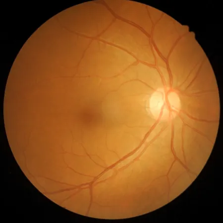
Could Your Retina Be at Risk? New Study Unveils Hidden Dangers!
2025-05-25
Author: Jia
New Insights into Retinal Atrophy Risk
A groundbreaking study from Sapienza University of Rome has uncovered alarming clues that specific optical coherence tomography (OCT) features might indicate a heightened risk of retinal atrophy (RA) for patients with neovascular age-related macular degeneration (nAMD). This research is crucial, as AMD stands as the leading cause of blindness in developed countries, with projections estimating a rise from 196 million affected individuals in 2020 to 288 million by 2040.
Understanding AMD and Its Impact
Age-related macular degeneration (AMD) is not just a health issue; it's a growing global crisis. Its prevalence varies significantly across different regions, hinting at underlying factors that aren't merely demographic.
Dr. Oscar M. Gagliardi, a leading researcher in the study, emphasizes the importance of identifying reliable indicators of AMD progression. "In our study, we sought to pinpoint OCT features that could predict RA in nAMD patients treated with aflibercept," he explains.
Study Breakdown: Who Were the Participants?
In this retrospective cohort study, researchers probed 1,085 clinical records. Out of these, 703 patients had undergone treatment. Ultimately, the analysis focused on 46 eligible eyes, with an average age of 78.37 years. The study presented a hopeful vision: while some eyes showed no sign of RA after five years, others met the criteria for RA, underscoring the diversity in patient outcomes.
Key Findings: What the Data Reveals
The study revealed a stark contrast in visual acuity outcomes between atrophic and non-atrophic groups five years post-diagnosis. At the beginning, there were no significant differences in best-corrected visual acuity (BCVA), but after five years, the atrophic group faced a dramatically poorer visual outcome. Meanwhile, the non-atrophic group experienced notable improvements in their vision.
Critical OCT Signals to Monitor
The study identified several OCT features linked to RA, such as type 2 macular neovascularization, reductions in the outer nuclear layer (ONL), and decreased central foveal thickness (CFT). Eyes that developed RA exhibited a thinner ONL and more intraretinal fluid at diagnosis.
Looking Ahead: Limitations and Future Directions
While the findings are promising, Gagliardi and his team caution against relying solely on OCT for RA assessments. They recommend incorporating various imaging methods in future studies to gain a more comprehensive understanding of AMD progression.
As the number of AMD cases continues to rise, ongoing research and innovative treatment strategies will be vital in combating this sight-threatening condition.


 Brasil (PT)
 Canada (EN)
 Chile (ES)
 Česko (CS)
 대한민국 (KO)
 España (ES)
 France (FR)
 Hong Kong (EN)
 Italia (IT)
 日本 (JA)
 Magyarország (HU)
 Norge (NO)
 Polska (PL)
 Schweiz (DE)
 Singapore (EN)
 Sverige (SV)
 Suomi (FI)
 Türkiye (TR)
 الإمارات العربية المتحدة (AR)