Breakthrough! Scientists Create Mini Amniotic Sacs in the Lab Using Stem Cells
2025-07-11
Author: Wei
A Revolutionary Lab Model for Human Development
In a groundbreaking achievement, researchers have successfully developed an advanced laboratory model that mimics the human amniotic sac during the crucial first weeks of embryonic development. This innovative model, crafted from stem cells, promises to unlock new frontiers in our understanding of human growth and may offer game-changing applications in various medical treatments, from healing burns to reconstructing corneas.
Understanding the Amniotic Sac's Vital Role
The amniotic sac, often described as a protective balloon filled with fluid, plays a vital role by cushioning the developing embryo. The liquid inside is thought to be essential for healthy growth. However, researching this significant interaction presents ethical challenges and logistical hurdles when studying human embryos directly.
A Major Advancement in Lab Models
Previous attempts at simulating the amniotic sac were limited, as they couldn’t replicate the complex two-layer structure and had short lifespans. In contrast, the new model, dubbed post-gastrulation amnioids (PGAs), thrives for at least three months and reaches the developmental stage of a month-old amniotic sac, growing up to an impressive inch in size.
Self-Organization: The Key to Success
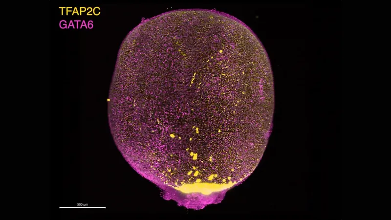
Leading the research, Silvia Santos from the Francis Crick Institute, explained that these little structures, resembling golf balls, formed through a remarkable self-organization process. Utilizing embryonic stem cells, the team employed a strategic signaling method that allowed the cells to communicate and assemble into the desired two-layer model.
The Influence of GATA3 Revealed
In their quest to decode the secrets of the amniotic sac, researchers identified a pivotal gene, GATA3, responsible for directing cells to develop into PGAs. This gene acts as a transcription factor, activating crucial signals that facilitate cell specialization.
Transforming Surrounding Cells
In an intriguing experiment, the PGAs were placed alongside unspecialized stem cells. Remarkably, these initial cells transformed into various "extraembryonic" cell types, showcasing the amniotic sac’s ability to influence nearby cells.
Endless Potential for Medical Advancements
The implications of this research are vast. With antimicrobial and anti-inflammatory properties, PGAs could become a reliable source for transplant tissues used in critical medical treatments. Yi Zheng, a biomedical engineering expert, suggested that converting patient-specific induced pluripotent stem cells into PGAs might optimize treatment applications.
Addressing Congenital Disorders
Furthermore, understanding the amniotic sac's structure through PGAs may shed light on congenital disorders linked to its size and contents, potentially paving the way for better preventive strategies.
Santos expressed her enthusiasm: "I'm extremely excited about the potential of these little structures," underlining the bright future that lies ahead in the realm of developmental biology and regenerative medicine.


 Brasil (PT)
 Canada (EN)
 Chile (ES)
 Česko (CS)
 대한민국 (KO)
 España (ES)
 France (FR)
 Hong Kong (EN)
 Italia (IT)
 日本 (JA)
 Magyarország (HU)
 Norge (NO)
 Polska (PL)
 Schweiz (DE)
 Singapore (EN)
 Sverige (SV)
 Suomi (FI)
 Türkiye (TR)
 الإمارات العربية المتحدة (AR)