Breakthrough in Terahertz Imaging Technology Reveals Cochlear Secrets and Revolutionizes Hearing Loss Diagnosis!
2025-04-02
Author: Li
A stunning advancement in imaging technology could change the way we diagnose hearing loss and other ear-related conditions. Researchers have harnessed the power of terahertz imaging to visualize the intricate internal structures of the cochlea at a remarkable micron-level resolution—for the first time ever!
A collaborative effort from a multi-institutional team has led to the development of an innovative 3D terahertz near-field imaging technique. This method provides exceptionally detailed images, enabling accurate three-dimensional reconstructions of the cochlea, the vital organ responsible for converting sound waves into neural signals.
The Science Behind Terahertz Imaging
Terahertz radiation occupies a unique place in the electromagnetic spectrum, bridging the gap between microwaves and mid-infrared light. Its low-energy nature makes terahertz imaging a safe option for biological tissues, as it does not harm them. Moreover, it is adept at penetrating bone while being sensitive to cellular structures and hydration levels.
Associate Professor Kazunori Serita from Waseda University in Japan, who leads the research team, elaborates on the significance of this technology: 'Conventional imaging methods often struggle to capture the fine details of the cochlea. Our new 3D imaging technique allows us to visualize small, critical structures inside this organ without any damage.'
The implications of this technology are profound. Serita notes, 'This method could provide innovative ways to diagnose ear diseases that have remained elusive so far, potentially enabling the on-site diagnosis of conditions like sensorineural hearing loss and other ear disorders. Early detection of hearing impairments could lead to earlier treatment options and significantly better outcomes for patients.'
The Technical Breakthrough
Serita's quest to develop this groundbreaking technique was inspired by discussions about cochlear measurement challenges with Associate Professor Takeshi Fujita from Kobe University. Together, they explored the possibilities of utilizing terahertz imaging to overcome these hurdles.
Unlike traditional imaging techniques that rely on bulky lenses limited by size, the researchers found a way to dispense with these constraints. They employed a nonlinear optical crystal to create terahertz light from a minuscule source, achieving a beam diameter of just 20 microns. This advance allowed them to successfully image the tiny internal structures of the cochlea without causing damage.
As Serita explains, 'Our pivotal innovation was generating terahertz waves from 1,560-nm near-infrared light. This was critical for achieving high-resolution imaging of the cochlea's internal anatomy.'
Successful Pilot Experiments
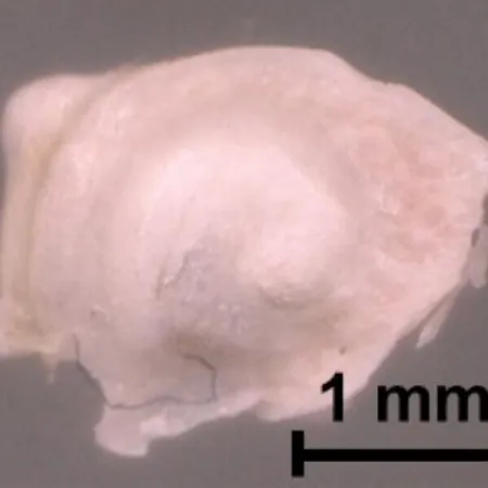
The team conducted tests using extracted and dried mouse cochlear samples—one empty and the other filled with a reflective metal—to confirm that terahertz waves could penetrate the cochlea effectively. They observed distinct differences between the two samples, which affirmed the technology's capability.
After confirming the approach, researchers utilized unsupervised learning algorithms to extract and visualize essential internal structural information from 2D terahertz time-domain images. They also succeeded in achieving 3D terahertz time-of-flight imaging, which revealed the intricate spiral structure of the cochlear duct.
Future Developments
Looking ahead, the research team aims to implement this imaging technique in a more natural biological setting. Given the cochlea's deep location within the ear and the surrounding lymphatic fluid, they plan to miniaturize their system for insertion through the ear canal. Additionally, they're developing stronger terahertz sources to reach deeper structures.
Once this terahertz imaging technology is refined and compacted, it holds the promise of being integrated into endoscopes and otoscopes, paving the way for non-invasive in vivo imaging. This could revolutionize cochlear diagnostics and might also hold potential for early cancer detection in various organs.
The Next Frontier in Ear Health
As researchers continue to push the boundaries of this innovative imaging technology, the future could look brighter for those affected by hearing loss and other ear-related health issues. With early detection and improved diagnostic capabilities, patients may soon benefit from targeted interventions and enhanced quality of life. Stay tuned, as the future of ear health is about to be transformed!


 Brasil (PT)
 Canada (EN)
 Chile (ES)
 Česko (CS)
 대한민국 (KO)
 España (ES)
 France (FR)
 Hong Kong (EN)
 Italia (IT)
 日本 (JA)
 Magyarország (HU)
 Norge (NO)
 Polska (PL)
 Schweiz (DE)
 Singapore (EN)
 Sverige (SV)
 Suomi (FI)
 Türkiye (TR)
 الإمارات العربية المتحدة (AR)