Breakthrough in Breast Imaging: The Painless, Precision Method Set to Revolutionize Cancer Detection!
2025-06-30
Author: Sarah
A Game-Changer in Breast Imaging
In a groundbreaking development, a team led by Caltech has unveiled a revolutionary breast imaging technique that is safe, effective, and completely painless. This innovative approach harnesses machine learning to differentiate between healthy and suspicious tissue, outperforming traditional methods in recent tests on patients.
The Limitations of Traditional Techniques
For years, X-ray mammography has been the oracle of breast cancer screening. While it has played a significant role in reducing cancer deaths, it is not without its flaws: exposure to ionizing radiation, painful squeezing of breast tissue, and a high rate of false positives, especially in dense breast tissue, complicate matters. Other imaging options like ultrasound and MRI also have their drawbacks—ultrasound's accuracy hinges on the operator's skill, and MRI is time-consuming and costly, often restricting it for claustrophobic patients or those with certain implants.
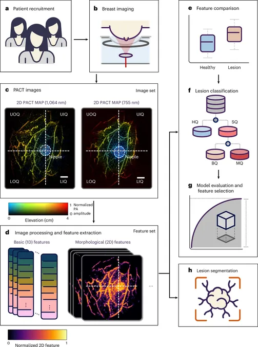
Introducing PACT: The Future of Breast Imaging
Enter the game-changing technology: Photoacoustic Computed Tomography (PACT), meticulously developed over the past two decades by Professor Lihong Wang and his team. This innovative method provides a viable alternative to conventional imaging without discomfort or steep costs, allowing for a quick scan that identifies tumors in just 15 seconds.
Clinical Triumphs
In collaboration with researchers from the City of Hope Comprehensive Cancer Center, PACT has shown exceptional results in trials involving 39 patients, effectively distinguishing between malignant and benign growths with accuracy comparable to mammography and MRI.
Behind the Science: How PACT Works
So, how does PACT work its magic? By employing a near-infrared laser pulse that penetrates breast tissue, the system detects molecular vibrations—specifically from hemoglobin. This unique combination of light and sound enables high-resolution imagery, allowing for precise visualization of internal structures, down to a quarter-millimeter at depths of 4 centimeters. PACT’s remarkable ability to identify functional differences in tissue helps in the early detection of diseases, making it a potent ally in the fight against breast cancer.
Pioneering AI in Medical Imaging
The integration of artificial intelligence (AI) has significantly amplified PACT's capabilities. The system has been trained to discern subtle tissue variations, enhancing its proficiency in detecting abnormalities that might escape the human eye—potentially catching issues before they become critical.
A Comfortable Patient Experience
During a PACT scan, patients lie comfortably face down on a specialized table that cradles one breast at a time. The rapid scan process means patients only need to hold their breath briefly, making the experience as convenient as it is innovative.
Looking Ahead: The Vision for PACT
As Professor Wang puts it, this accomplishment is the culmination of decades of dedication to improving breast cancer detection. The goal? To make PACT a clinical tool that revolutionizes breast imaging, offering safer, faster, and more efficient screenings that could save countless lives without the worry of radiation or allergic reactions.


 Brasil (PT)
 Canada (EN)
 Chile (ES)
 Česko (CS)
 대한민국 (KO)
 España (ES)
 France (FR)
 Hong Kong (EN)
 Italia (IT)
 日本 (JA)
 Magyarország (HU)
 Norge (NO)
 Polska (PL)
 Schweiz (DE)
 Singapore (EN)
 Sverige (SV)
 Suomi (FI)
 Türkiye (TR)
 الإمارات العربية المتحدة (AR)